VSports最新版本 - Skip to main page content
U.S. flag

An official website of the United States government

Dot gov

The . gov means it’s official. Federal government websites often end in . gov or . mil VSports app下载. Before sharing sensitive information, make sure you’re on a federal government site. .

Https

The site is secure V体育官网. The https:// ensures that you are connecting to the official website and that any information you provide is encrypted and transmitted securely. .

. 2012 Sep 7;337(6099):1222-5.
doi: 10.1126/science.1219379.

Rad51 is an accessory factor for Dmc1-mediated joint molecule formation during meiosis

Affiliations

Rad51 is an accessory factor for Dmc1-mediated joint molecule formation during meiosis

"VSports注册入口" Veronica Cloud et al. Science. .

Abstract

Meiotic recombination in budding yeast requires two RecA-related proteins, Rad51 and Dmc1, both of which form filaments on DNA capable of directing homology search and catalyzing formation of homologous joint molecules (JMs) and strand exchange VSports手机版. With use of a separation-of-function mutant form of Rad51 that retains filament-forming but not JM-forming activity, we show that the JM activity of Rad51 is fully dispensable for meiotic recombination. The corresponding mutation in Dmc1 causes a profound recombination defect, demonstrating Dmc1's JM activity alone is responsible for meiotic recombination. We further provide biochemical evidence that Rad51 acts with Mei5-Sae3 as a Dmc1 accessory factor. Thus, Rad51 is a multifunctional protein that catalyzes recombination directly in mitosis and indirectly, via Dmc1, during meiosis. .


"VSports app下载" Figures

Fig. 1
Fig. 1
Rad51-II3A binds DNA to form nucleoprotein filaments. (A) Rad51-WT (closed circle) and Rad51-II3A (open-square) bind oligo-dT as revealed by fluorescence polarization. Error bars represent SEM, N = 4. The apparent affinity of both proteins is about 300 nM. (B) EMSA analysis of DNA binding. Protein (at 0, 1, 2, 4, and 6 μM) binds M13mp18 ssDNA (22 μM nucleotides) and linear pBluescript dsDNA (8 μM base pairs). The inverted images of agarose gels are presented. The percentage of DNA in Rad51-DNA complexes is plotted. SEM, N = 6 for ssDNA, N = 3 for dsDNA. (C) Rad51-WT and Rad51-II3A proteins form filaments on a 1000-nucleotide fragment of ØX174 ssDNA. (D) ssDNA-Rad51-WT but not ssDNA-Rad51-II3A filaments bind 32P-pBluescript dsDNA (18 μM base pairs). Filament concentrations = 0, 1.5, 2.9, 4.4, 5.8, and 7.3 μM protomer (at 3nt/protomer). Phorphorimage of radioactive EMSA gel is presented. For plotted data, N= 3.
Fig. 2
Fig. 2
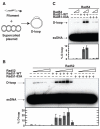
ScRad51-II3A mutant protein is defective in D-loop activity. (A) Schematic of D-loop assay. (B,C) D-loop formation by Rad51-WT or Rad51-II3A (1.2 μM) was examined with or without ScRad52 (0.3, 0.6, 1.2, 2.4 and 4.8 μM) in (B); with or without ScRad54 (0.2 and 0.4 μM) in (C). Rad51 was pre-incubated with either Rad52 or Rad54, followed by incubation with 32P-labeled 90-mer ssDNA (3.6 μM nucleotides) at 37 °C. D-loops were generated upon the addition of plasmid (22 μM base pairs) to the mixture. Error bars represent SEM, N=3.
Fig. 3
Fig. 3
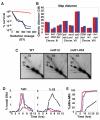
Phenoptypic analysis of rad51-II3A mutants. (A) Cell survival as a function of γ ray dose for WT (DKB3507, red), rad51-II3A (DKB3690, blue), and rad51Δ (DKB3048, black). (B) Genetic map distances (with standard error) from diploids WT (NHY1848, red) and rad51-II3A (DKB4005, blue). (C) 2-D gel analysis of IH JMs at HIS4::LEU2. Representative images showing peak time points are presented. The positions IH and IS JM species are shown (21). (D) IH and IS as a percentage of total DNA: WT (red), rad51-II3A (blue), rad51Δ (black). (E) Timing of meiotic divisions % MI ± MII = number of cells with >1 DAPI staining body over the total # of cells X100.
Fig. 4
Fig. 4
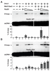
Rad51 stimulates Dmc1’s D-loop activity. Dmc1 (1 μM) was pre-incubated with or without Rad51-WT (A) or Rad51-II3A (B) (0.5, 0.1, and 0.02 μM), Mei5-Sae3 (0.5 μM), and 32P-labeled 90-mer ssDNA (3.6 μM nucleotides). Plasmid pRS306 (22 μM base pairs) was then added to initiate D-loop formation. Error bars represent SEM, N=4.

References

    1. Bishop DK, Park D, Xu L, Kleckner N. Cell. 1992;69:439. - PubMed
    1. Shinohara A, Ogawa H, Ogawa T. Cell. 1992;69:457. - PubMed
    1. Schwacha A, Kleckner N. Cell. 1997 Sep 19;90:1123. - PubMed
    1. Sheridan S, Bishop DK. Genes Dev. 2006 Jul 1;20:1685. - PubMed (V体育2025版)
    1. Tsubouchi H, Roeder GS. Genes Dev. 2006 Jul 1;20:1766. - PMC - PubMed

Publication types

"V体育平台登录" MeSH terms