Skip to main page content
U.S. flag

An official website of the United States government

Dot gov

The . gov means it’s official. Federal government websites often end in . gov or VSports app下载. mil. Before sharing sensitive information, make sure you’re on a federal government site. .

Https

The site is secure. The https:// ensures that you are connecting to the official website and that any information you provide is encrypted and transmitted securely V体育官网. .

. 2012 Oct 15;11(20):3810-27.
doi: 10.4161/cc.22022. Epub 2012 Sep 5.

Phospho-ΔNp63α/SREBF1 protein interactions: bridging cell metabolism and cisplatin chemoresistance

Affiliations

Phospho-ΔNp63α/SREBF1 protein interactions: bridging cell metabolism and cisplatin chemoresistance (VSports)

Yiping Huang et al. Cell Cycle. .

Abstract

Tumor protein (TP)-p53 family members (TP63, TP63 and TP73) are guardians of the genome and key players in orchestrating the cellular response to cisplatin treatment VSports手机版. Cisplatin-induced phosphorylation of ΔNp63α was shown to have a role in regulating intracellular ΔNp63α protein levels. We previously found that squamous cell carcinoma (SCC) cells exposed to cisplatin displayed the ATM-dependent phosphorylation of ΔNp63α (p-ΔNp63α), which is critical for the transcriptional regulation of specific downstream mRNAs and microRNAs and is likely to underlie the chemoresistance of SCC cells. However, SCC cells expressing non-p-ΔNp63α became more cisplatin-resistant. We also found that p-ΔNp63α forms complexes with a number of proteins involved in cell death response through regulation of cell cycle arrest, apoptosis, autophagy, RNA splicing and chromatin modifications. Here, we showed that p-ΔNp63α induced ARG1, GAPDH, and CPT2 gene transcription in cisplatin-sensitive SCC cells, while non-p-ΔNp63α increased a transcription of CAD, G6PD and FASN genes in cisplatin-resistant SCC cells. We report that the p-ΔNp63α-dependent regulatory mechanisms implicated in the modulation of plethora of pathways, including amino acid, carbohydrate, lipid and nucleotide metabolisms, thereby affect tumor cell response to cisplatin-induced cell death, suggesting that the ATM-dependent ΔNp63α pathway plays a role in the resistance of tumor cells to platinum therapy. .


V体育官网 - Figures
None
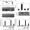
Figure 1. P-ΔNp63α induces the expression of ARG1 in SCC-11 cells upon cisplatin exposure. (A) Metabolite reaction of arginase 1 (ARG1). Grey arrow indicates the increase in SCC-11 cells, while black arrow indicates the decrease in SCC-11M cells. (B, upper panel) qPCR expression analysis of ARG1 mRNA obtained from SCC-11 and SCC-11M cells treated with control medium (Con) and 10μg/ml cisplatin (CIS). The values from SCC-11 cells treated with control medium designated as 1. The qPCR values presented as relative units (RU) (B, lower panel) Immunoblot analysis of ARG1 in SCC-11 and SCC-11M cells treated with control medium (Con) or cisplatin (CIS). Loading was normalized by β-actin expression. (C) ChIP assay of the binding of p-ΔNp63α and SREBF1 to the ARG1 promoter (enrichment) in SCC-11 cells exposed to control medium (Con) and cisplatin (CIS). (D) Quantitative analysis of enrichment (ChIP/Input ratio) of the ARG1 promoter with the p-ΔNp63α and SREBF1 transcription factors in SCC-11 cells treated with cisplatin. ChiP/Input ratio values presented as relative units (RU). SCC-11 cells were transfected with the scrambled and SREBF1 siRNA and analyzed for ChIP/Input enrichment. (E) Luciferase reporter assay of the ARG1 promoter in SCC-11 and SCC-11M cells treated with control medium (Con) and cisplatin (CIS). SCC-11 cells were transfected with the scrambled and SREBF1 siRNA and analyzed for the ARG1 promoter-driven luciferase activity presented as relative units (RU). The values from SCC-11 cells treated with control medium designated as 1. All experiments were performed in triplicates. p < 0.05.
None
Figure 4. P-ΔNp63α induces the expression of CPT2 in SCC-11 cells upon cisplatin exposure. (A) Metabolite reaction of carnitine palmitoyltransferase 2 (CPT2). Grey arrow indicates the increase in SCC-11 cells, while black arrow indicates the decrease in SCC-11M cells. (B, upper panel) qPCR expression analysis of CPT2 mRNA obtained from SCC-11 and SCC-11M cells treated with control medium (Con) and 10 μg/ml cisplatin (CIS). The values from SCC-11 cells treated with control medium designated as 1. The qPCR values presented as relative units (RU). (B, lower panel) Immunoblot analysis of CPT2 in SCC-11 and SCC-11M cells treated with control medium (Con) or cisplatin (CIS). Loading was normalized by β-actin expression. (C) ChIP assay of the binding of p-ΔNp63α and SREBF1 to the CPT2 promoter (enrichment) in SCC-11 cells exposed to control medium (Con) and cisplatin (CIS). (D) Quantitative analysis of enrichment (ChIP/Input ratio) of the CPT2 promoter with the p-ΔNp63α and SREBF1 transcription factors in SCC-11 cells treated with cisplatin. ChiP/Input ratio values presented as relative units (RU). SCC-11 cells were transfected with the scrambled and SREBF1 siRNA and analyzed for ChIP/Input enrichment. (E) Luciferase reporter assay of the CPT2 promoter in SCC-11 and SCC-11M cells treated with control medium (Con) and cisplatin (CIS). SCC-11 cells were transfected with the scrambled and SREBF1 siRNA and analyzed for the CPT2 promoter-driven luciferase activity presented as relative units (RU). The values from SCC-11 cells treated with control medium designated as 1. All experiments were performed in triplicates. p < 0.05.
None
Figure 2. P-ΔNp63α induces the expression of GAPDH in SCC-11 cells upon cisplatin exposure. (A) Metabolite reaction of glyceraldehyde-3-phosphate dehydrogenase. Grey arrow indicates the increase in SCC-11 cells, while black arrow indicates the decrease in SCC-11M cells. (B, upper panel) qPCR expression analysis of GAPDH mRNA obtained from SCC-11 and SCC-11M cells treated with control medium (Con) and 10 μg/ml cisplatin (CIS). The values from SCC-11 cells treated with control medium designated as 1. The qPCR values presented as relative units (RU). (B, lower panel) Immunoblot analysis of GAPDH in SCC-11 and SCC-11M cells treated with control medium (Con) or cisplatin (CIS). Loading was normalized by β-actin expression. (C) ChIP assay of the binding of p-ΔNp63α and SREBF1 to the GAPDH promoter (enrichment) in SCC-11 cells exposed to control medium (Con) and cisplatin (CIS). (D) Quantitative analysis of enrichment (ChIP/Input ratio) of the GAPDH promoter with the p-ΔNp63α and SREBF1 transcription factors in SCC-11 cells treated with cisplatin. ChiP/Input ratio values presented as relative units (RU). SCC-11 cells were transfected with the scrambled and SREBF1 siRNA and analyzed for ChIP/Input enrichment. (E) Luciferase reporter assay of the GAPDH promoter in SCC-11 and SCC-11M cells treated with control medium (Con) and cisplatin (CIS). SCC-11 cells were transfected with the scrambled and SREBF1 siRNA and analyzed for the GAPDH promoter-driven luciferase activity presented as relative units (RU). The values from SCC-11 cells treated with control medium designated as 1. All experiments were performed in triplicates. p < 0.05.
None
Figure 3. P-ΔNp63α induces the expression of PKM2 in SCC-11 cells upon cisplatin exposure. (A) Metabolite reaction of pyruvate kinase M2 (PKM2). Grey arrow indicates the increase in SCC-11 cells, while black arrow indicates the decrease in SCC-11M cells. (B, upper panel) qPCR expression analysis of PKM2 mRNA obtained from SCC-11 and SCC-11M cells treated with control medium (Con) and 10 μg/ml cisplatin (CIS). The values from SCC-11 cells treated with control medium designated as 1. The qPCR values presented as relative units (RU). (B, lower panel) Immunoblot analysis of PKM2 in SCC-11 and SCC-11M cells treated with control medium (Con) or cisplatin (CIS). Loading was normalized by β-actin expression. (C) ChIP assay of the binding of p-ΔNp63α and SREBF1 to the PKM2 promoter (enrichment) in SCC-11 cells exposed to control medium (Con) and cisplatin (CIS). (D) Quantitative analysis of enrichment (ChIP/Input ratio) of the PKM2 promoter with the p-ΔNp63α and SREBF1 transcription factors in SCC-11 cells treated with cisplatin. ChiP/Input ratio values presented as relative units (RU). SCC-11 cells were also transfected with the scrambled and SREBF1 siRNA and analyzed for ChIP/Input enrichment. (E) Luciferase reporter assay of the PKM2 promoter in SCC-11 and SCC-11M cells treated with control medium (Con) and cisplatin (CIS). SCC-11 cells were transfected with the scrambled and SREBF1 siRNA and analyzed for the PKM2 promoter-driven luciferase activity presented as relative units (RU). The values from SCC-11 cells treated with control medium designated as 1. All experiments were performed in triplicates. p < 0.05.
None
Figure 5. Non-p-ΔNp63α induces the expression of CAD in SCC-11M cells upon cisplatin exposure. (A) Metabolite reaction of carbamoyl-phosphate synthetase 2 (CAD). Grey arrow indicates the decrease in SCC-11 cells, while black arrow indicates the increase in SCC-11M cells. (B, upper panel) qPCR expression analysis of CAD mRNA obtained from SCC-11 and SCC-11M cells treated with control medium (Con) and 10 μg/ml cisplatin (CIS). The values from SCC-11 cells treated with control medium designated as 1. The qPCR values presented as relative units (RU). (B, lower panel) Immunoblot analysis of CAD in SCC-11 and SCC-11M cells treated with control medium (Con) or cisplatin (CIS). Loading was normalized by β-actin expression. (C) ChIP assay of the binding of p-ΔNp63α and E2F1 to the CAD promoter (enrichment) in SCC-11M cells exposed to control medium (Con) and cisplatin (CIS). (D) Quantitative analysis of enrichment (ChIP/Input ratio) of the CAD promoter with the p-ΔNp63α and E2F1 transcription factors in SCC-11M cells treated with cisplatin. ChiP/Input ratio values presented as relative units (RU). SCC-11M cells were transfected with the scrambled and E2F1 siRNA and analyzed for ChIP/Input enrichment. (E) Luciferase reporter assay of the CAD promoter in SCC-11 and SCC-11M cells treated with control medium (Con) and cisplatin (CIS). SCC-11M cells were transfected with the scrambled and E2F1 siRNA and analyzed for the CAD promoter-driven luciferase activity presented as relative units (RU). The values from SCC-11 cells treated with control medium designated as 1. All experiments were performed in triplicates. p < 0.05.
None
Figure 8. Non-p-ΔNp63α induces the expression of FASN in SCC-11M cells upon cisplatin exposure. (A) Metabolite reaction of fatty acid synthase (FASN). Grey arrow indicates the decrease in SCC-11 cells, while black arrow indicates the increase in SCC-11M cells. (B, upper panel) qPCR expression analysis of FASN mRNA obtained from SCC-11 and SCC-11M cells treated with control medium (Con) and 10 μg/ml cisplatin (CIS). The values from SCC-11 cells treated with control medium designated as 1. The qPCR values presented as relative units (RU). (B, lower panel) Immunoblot analysis of FASN in SCC-11 and SCC-11M cells treated with control medium (Con) or cisplatin (CIS). Loading was normalized by β-actin expression. (C) ChIP assay of the binding of p-ΔNp63α and E2F1 to the FASN promoter (enrichment) in SCC-11M cells exposed to control medium (Con) and cisplatin (CIS). (D) Quantitative analysis of enrichment (ChIP/Input ratio) of the FASN promoter with the p-ΔNp63α and E2F1 transcription factors in SCC-11M cells treated with cisplatin. ChiP/Input ratio values presented as relative units (RU). SCC-11M cells were transfected with the scrambled and E2F1 siRNA and analyzed for ChIP/Input enrichment. (E) Luciferase reporter assay of the FASN promoter in SCC-11 and SCC-11M cells treated with control medium (Con) and cisplatin (CIS). SCC-11M cells were transfected with the scrambled and E2F1 siRNA and analyzed for the FASN promoter-driven luciferase activity presented as relative units (RU). The values from SCC-11 cells treated with control medium designated as 1. All experiments were performed in triplicates. p < 0.05.
None
Figure 6. Non-p-ΔNp63α induces the expression of G6PD in SCC-11M cells upon cisplatin exposure. (A) Metabolite reaction of glucose-6-phosphate dehydrogenase (G6PD). Grey arrow indicates the decrease in SCC-11 cells, while black arrow indicates the increase in SCC-11M cells. (B, upper panel) qPCR expression analysis of G6PD mRNA obtained from SCC-11 and SCC-11M cells treated with control medium (Con) and 10μg/ml cisplatin (CIS). The values from SCC-11 cells treated with control medium designated as 1. The qPCR values presented as relative units (RU). (B, lower panel) Immunoblot analysis of G6PD in SCC-11 and SCC-11M cells treated with control medium (Con) or cisplatin (CIS). Loading was normalized by β-actin expression. (C) ChIP assay of the binding of p-ΔNp63α and E2F1 to the G6PD promoter (enrichment) in SCC-11M cells exposed to control medium (Con) and cisplatin (CIS). (D) Quantitative analysis of enrichment (ChIP/Input ratio) of the G6PD promoter with the p-ΔNp63α and E2F1 transcription factors in SCC-11M cells treated with cisplatin. ChiP/Input ratio values presented as relative units (RU). SCC-11M cells were transfected with the scrambled and E2F1 siRNA and analyzed for ChIP/Input enrichment. (E) Luciferase reporter assay of the G6PD promoter in SCC-11 and SCC-11M cells treated with control medium (Con) and cisplatin (CIS). SCC-11M cells were transfected with the scrambled and E2F1 siRNA and analyzed for the G6PD promoter-driven luciferase activity presented as relative units (RU). The values from SCC-11 cells treated with control medium designated as 1. All experiments were performed in triplicates. p < 0.05.
None
Figure 7. Non-p-ΔNp63α induces the expression of PFKFB3 in SCC-11M cells upon cisplatin exposure. (A) Metabolite reaction of fructose-2, 6-biphosphatase (PFKFB3). Grey arrow indicates the decrease in SCC-11 cells, while black arrow indicates the increase in SCC-11M cells. (B, upper panel) qPCR expression analysis of PFKFB3 mRNA obtained from SCC-11 and SCC-11M cells treated with control medium (Con) and 10 μg/ml cisplatin (CIS). The values from SCC-11 cells treated with control medium designated as 1. The qPCR values presented as relative units (RU). (B, lower panel) Immunoblot analysis of PFKFB3 in SCC-11 and SCC-11M cells treated with control medium (Con) or cisplatin (CIS). Loading was normalized by β-actin expression. (C) ChIP assay of the binding of p-ΔNp63α and E2F1 to the PFKFB3 promoter (enrichment) in SCC-11M cells exposed to control medium (Con) and cisplatin (CIS). (D) Quantitative analysis of enrichment (ChIP/Input ratio) of the PFKFB3 promoter with the p-ΔNp63α and E2F1 transcription factors in SCC-11M cells treated with cisplatin. ChiP/Input ratio values presented as relative units (RU). SCC-11M cells were transfected with the scrambled and E2F1 siRNA and analyzed for ChIP/Input enrichment. (E) Luciferase reporter assay of the PFKFB3 promoter in SCC-11 and SCC-11M cells treated with control medium (Con) and cisplatin (CIS). SCC-11M cells were transfected with the scrambled and E2F1 siRNA and analyzed for the PFKFB3 promoter-driven luciferase activity presented as relative units (RU). The values from SCC-11 cells treated with control medium designated as 1. All experiments were performed in triplicates. p < 0.05.
None
Figure 9. siRNA-dependent attenuation of sensitivity of SCC-11 cells and resistance of SCC-11M cells to cisplatin-induced cell death. SCC-11 cells were transfected with the scrambled and ARG1 siRNA (A), or the scrambled and CPT2 siRNA (B), while SCC-11M cells were transfected with the scrambled or G6PD siRNA (C) or the scrambled and FASN siRNA (D) for 32 h. Resulting cells were exposed to control medium (Con) or 10 μg/ml cisplatin (CIS) for 5 d, as indicated. Cell viability (MTT) assay was repeated three times. The bars are the mean ± SD of triplicate; p < 0.05.
None
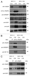
Figure 10. P-ΔNp63α/SREBF1 and non-p-ΔNp63α/E2F1 protein complexes formed in SCC cells upon cisplatin exposure. (A) Immunoblotting analysis of SCC-11 and SCC-11M cells exposed to control medium (Con) and 10 μg/ml cisplatin (CIS). Nuclear lysates were probed for expression of specific transcription factors with indicated antibodies. Levels of TBP served as loading controls. (B) Immunoprecipitation (IP) with anti-p-ΔNp63α antibody followed by immunoblotting with anti-SREBF1 and NF-Y antibodies. (C) IP with anti-ΔNp63 antibody followed by immunoblotting with anti-E2F1 and anti-p53 antibodies.

References

    1. Helmbach H, Kern MA, Rossmann E, Renz K, Kissel C, Gschwendt B, et al. Drug resistance towards etoposide and cisplatin in human melanoma cells is associated with drug-dependent apoptosis deficiency. J Invest Dermatol. 2002;118:923–32. doi: 10.1046/j.1523-1747.2002.01786.x. - DOI - PubMed
    1. Zangen R, Ratovitski EA, Sidransky D. DeltaNp63α levels correlate with clinical tumor response to cisplatin. Cell Cycle. 2005;4:1313–5. doi: 10.4161/cc.4.10.2066. - "VSports app下载" DOI - PubMed
    1. Kelland L. The resurgence of platinum-based cancer chemotherapy. Nat Rev Cancer. 2007;7:573–84. doi: 10.1038/nrc2167. - V体育2025版 - DOI - PubMed
    1. Galluzzi L, Senovilla L, Vitale I, Michels J, Martins I, Kepp O, et al. Molecular mechanisms of cisplatin resistance. Oncogene. 2012;31:1869–83. doi: 10.1038/onc.2011.384. - V体育官网入口 - DOI - PubMed
    1. Flores ER, Tsai KY, Crowley D, Sengupta S, Yang A, McKeon F, et al. Tsai K, Crowley D, Sengupta S, Yang A, McKeon F, Jacks T. P63 and p73 are required for TP53-dependent apoptosis in response to DNA damage. Nature. 2002;416:560–4. doi: 10.1038/416560a. - DOI - PubMed

MeSH terms