Skip to main page content
U.S. flag

An official website of the United States government

Dot gov

The . gov means it’s official. Federal government websites often end in . gov or VSports app下载. mil. Before sharing sensitive information, make sure you’re on a federal government site. .

Https

The site is secure V体育官网. The https:// ensures that you are connecting to the official website and that any information you provide is encrypted and transmitted securely. .

. 2012 Nov 1;72(21):5494-504.
doi: 10.1158/0008-5472.CAN-11-3993. Epub 2012 Aug 31.

VSports最新版本 - IKK-ε coordinates invasion and metastasis of ovarian cancer

Affiliations

IKK-ε coordinates invasion and metastasis of ovarian cancer

Sarah Hsu et al. Cancer Res. .

Abstract

Inhibitor of IκB kinases (IKK) are key regulators of NF-κB signaling. Three IKK isoforms-α, β, and ε-have been linked to oncogenesis, yet the precise components of NF-κB signaling in ovarian cancer have not yet been dissected. We surveyed 120 ovarian cancer specimens for IKK-ε expression. Notably, cytoplasmic expression was elevated in metastatic lesions relative to primary tumors (P = 0. 03) VSports手机版. Therefore, we hypothesized that IKK-ε drives ovarian cancer metastasis. IKK-ε was identified previously as a breast cancer oncogene and was associated with poor clinical outcome in ovarian cancer. We now define an ovarian cancer-specific IKK-ε-regulated gene expression signature using stably expressed short hairpin RNA targeting IKK-ε. Pathway analysis of the signature indicated that IKK-ε regulates expression of genes involved in cell motility and inflammation. We further showed that IKK-ε depletion in metastatic ovarian cancer cell lines decreased growth, adhesion, and invasion. Consistently, human xenografts depleted of IKK-ε in mice showed decreased aggressiveness, whereas overexpression of IKK-ε in a less invasive ovarian cancer cell line increased metastasis in vivo. Taken together, these data provide evidence that IKK-ε is a key coordinator of invasion and metastasis programs in ovarian cancer. Inhibition of IKK-ε signaling thus emerges as a viable therapeutic strategy in women whose ovarian cancer shows aberrant activation of this pathway. .


Conflict of interest statement

There are no conflicts to disclose

Figures
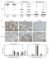
Figure 1
Figure 1. IKKε is higher in metastatic ovarian cancers compared to primary tumors
(A) Ovarian cancer tissue specimens were analyzed for relative expression of IKK isoforms – α, β, ε, - in primary and metastatic tumors. Expression (IHC score) is based on percent of cells showing expression of each protein (7). (B) Representative cases are shown at a 200 X magnification. (C) TCGA datasets were examined for expression of each IKK isoform (left) and copy number for the IKKα, IKKβ and IKKε loci (right) in ovarian cancer cases compared to non-cancer specimens. Relative expression is shown as log2 ratio of tumor to normal.
Figure 2
Figure 2. IKKε shRNAs specifically knock down its expression in ovarian cancer cells
(A) IKKε protein was highly expressed by Western blot in the majority of ovarian cancer cell lines. (B) Three shRNA constructs were tested for specificity towards IKKε, compared to IKKα or IKKβ, in cell lines Ovcar5, Ovcar8 and HeyA8. Knockdown of IKKε was measured after 1 week selection in puromycin, and 5 weeks after maintenance in medium without puromycin.
Figure 3
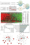
Figure 3. IKKε regulates expression of genes involved in cellular motility and inflammation
(A) Schematic of workflow for determining the ovarian cancer-specific IKKε gene signature. First, 278 genes were experimentally determined by depleting IKKε with 2 different shRNA constructs in Ovcar5 cells. In parallel, 456 genes were found to be associated with IKKε over-expression in 283 publicly available ovarian cancer gene expression profiles (GSE9899). The intersection of these two sets identified 17 genes. (B) The relative expression of these 17 genes in 283 ovarian cancer patient specimens is ranked by average expression of the 17 genes. Correlation of each gene with IKKε expression is also noted. (C) The 17-gene expression average was calculated for each of the 6 previously published subgroups of ovarian cancer (12). Log2 average was median-centered for the entire dataset. (D, E) The 17 genes were analyzed by Ingenuity Pathway Analysis. The most significantly related pathway was a cell-motility network, centered on the NF-κB complex.
Figure 4
Figure 4. IKKε modestly affects ovarian cancer growth and invasion in vitro
(A–C) Ovarian cancer cell lines Ovcar 5, Ovcar8, and HeyA8 were quantified by flow cytometry for GFP expression (co-expressed with shRNA construct) for 12–17 days after selection for IKKε depletion. Measurements are normalized to day 0 for each cell line. (D) Invasion through basement membrane was measured after depletion of IKKε, and normalized to control shRNA for each cell line. Error bars represent S.E.
Figure 5
Figure 5. IKKε promotes tumor growth, invasion and metastasis in vivo
(A) Persistent integration of the shRNA construct in Ovcar8 cells over the time course of mouse xenograft experiments was confirmed by Western blot of IKKε protein (2.5 weeks) and co-expressed GFP (5 weeks). β-tubulin was a loading control. (B) Organ weight was measured in mice after necropsy. Shown are the average organ weights for diaphragm, pancreas and liver (Ovcar5 xenografts, n=9 each). Organ weight is expressed as percent body weight, increased over organs from non-xenograft controls (PBS injected intraperitoneally). Error bars represent S.E. (C) Gross anatomical examination of the abdominal cavity showed decreased adhesion to external organ surfaces in IKKε-depleted Ovcar8 xenografts (left panels). Tumor adherence to pancreas and diaphragm also appeared decreased (right panels). (D) Quantification of xenograft invasion into secondary organs showed decreased invasiveness of Ovcar5 cells depleted of IKKε by either of the shRNA constructs. Shown is the number of animals with tumor present in each organ. (E) Immunohistochemical analysis of tissues exhibiting Ovcar5 xenografts, stained for GFP as a marker of shRNA construct integration. GFP protein is shown by the brown immunohistochemical stain, at 10X magnification.
Figure 6
Figure 6. IKKε overexpression promotes ovarian cancer metastasis
(A) Integration of the IKKε expression constructs was confirmed in Caov3 cells after 2 weeks in the presence of selection agent (G418) and after maintaining the culture without selection for 4 weeks (timecourse of xenograft experiments) as confirmed by Western blot of IKKε protein. GAPDH was a loading control. (B) Invasion through basement membrane was measured with overexpression of two IKKε constructs, and normalized to XTT viability for each cell line. (C) Shown is the relative expression of 7 IKKε-related metastasis-related genes. Expression was normalized to GAPDH and shown as fold change from cells transduced with empty vector. (D) Mice (n=6 each) were injected with Caov3 cell line engineered to express one of two different IKKε expression constructs or control. Weights were measured weekly. (E) Xenograft present in the peritoneal cavity or omental fat is denoted as “primary;” xenografts in other abdominal organs (liver, diaphragm, pancreas, other sites) are noted as “secondary.” Shown is the percent of mice with tumor in each of these locations. (F) Photomicrographs of representative tissue sections demonstrating superficial implantation of xenograft (top) or invasion into organ parenchyma (middle and bottom).

References

    1. Pignata S, Cannella L, Leopardo D, Pisano C, Bruni GS, Facchini G. Chemotherapy in epithelial ovarian cancer. Cancer Lett. 2011;303:73–83. - PubMed
    1. Annunziata CM, Azad N, Dhamoon AS, Whiteley G, Kohn EC. Ovarian cancer in the proteomics era. Int J Gynecol Cancer. 2008;18 (Suppl 1):1–6. - PubMed
    1. Martin LP, Schilder RJ. Management of recurrent ovarian carcinoma: current status and future directions. Semin Oncol. 2009;36:112–125. - "V体育平台登录" PubMed
    1. Bonome T, Levine DA, Shih J, Randonovich M, Pise-Masison CA, Bogomolniy F, et al. A gene signature predicting for survival in suboptimally debulked patients with ovarian cancer. Cancer Res. 2008;68:5478–86. - PMC - PubMed
    1. TCGA. Integrated genomic analyses of ovarian carcinoma. Nature. 2011;474:609–15. - PMC - PubMed

Publication types

MeSH terms