Skip to main page content
U.S. flag

An official website of the United States government

Dot gov

The . gov means it’s official VSports app下载. Federal government websites often end in . gov or . mil. Before sharing sensitive information, make sure you’re on a federal government site. .

Https

The site is secure. The https:// ensures that you are connecting to the official website and that any information you provide is encrypted and transmitted securely. V体育官网.

. 2012 Sep 11;109(37):15048-53.
doi: 10.1073/pnas.1203085109. Epub 2012 Aug 27.

Regulation of beta catenin signaling and parathyroid hormone anabolic effects in bone by the matricellular protein periostin

Affiliations

Regulation of beta catenin signaling and parathyroid hormone anabolic effects in bone by the matricellular protein periostin

Nicolas Bonnet et al. Proc Natl Acad Sci U S A. .

Abstract

Periostin (Postn) is a matricellular protein preferentially expressed by osteocytes and periosteal osteoblasts in response to mechanical stimulation and parathyroid hormone (PTH). Whether and how periostin expression influences bone anabolism, however, remains unknown. We investigated the skeletal response of adult Postn(-/-) and Postn(+/+) mice to intermittent PTH. Compared with Postn(+/+), Postn(-/-) mice had a lower bone mass, cortical bone volume, and strength response to PTH. PTH-stimulated bone-forming indices were all significantly lower in Postn(-/-) mice, particularly at the periosteum. Furthermore, in vivo stimulation of Wnt-β-catenin signaling by PTH, as evaluated in TOPGAL reporter mice, was inhibited in the absence of periostin (TOPGAL;Postn(-/-) mice). PTH stimulated periostin and inhibited MEF2C and sclerostin (Sost) expression in bone and osteoblasts in vitro. Recombinant periostin also suppressed Sost expression, which was mediated through the integrin αVβ3 receptor, whereas periostin-blocking antibody prevented inhibition of MEF2C and Sost by PTH. In turn, administration of a Sost-blocking antiboby partially restored the PTH-mediated increase in bone mass in Postn(-/-) mice. In addition, primary osteoblasts from Postn(-/-) mice showed a lower proliferation, mineralization, and migration, both spontaneously and in response to PTH. Osteoblastic gene expression levels confirmed a defect of Postn(-/-) osteoblast differentiation with and without PTH, as well as an increased osteoblast apoptosis in the absence of periostin VSports手机版. These data elucidate the complex role of periostin on bone anabolism, through the regulation of Sost, Wnt-β-catenin signaling, and osteoblast differentiation. .


Conflict of interest statement

The authors declare no conflict of interest.

Figures
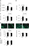
Fig. 1.
Fig. 1.
Effects of iPTH on bone in Postn−/− and Postn+/+ mice. (A) Femur bone BMD gain. (B) Trabecular BV/TV of the distal femur. (C) CtBV and CtTh at midshaft femur. (D) Fluorescent calcein labels on periosteal and endosteal surfaces. (E) Biomechanical properties of the cortical tibia. (F) Body weight gain. *P < 0.05, **P < 0.01, ***P < 0.001 significant difference vs. vehicle. Bars show mean (± SEM). Closed bars, iPTH; open bars, vehicle.
Fig. 2.
Fig. 2.
Postn and Sost protein expression in response to iPTH. (A) Immunohistochemical staining of Postn in longitudinal sections of the proximal tibia of Postn+/+ mice 24 h after PTH treatment shows increased Postn expression at the periosteum (Ps), but not at the endocortical surfaces (Ec). (B) Immunohistochemical staining of Sost expression in tibia midshaft sections showing decreased expression 24 h after PTH treatment in Postn+/+ but not Postn−/− mice. Note the presence of Sost into tin osteocyte canaliculae (white arrow) that disappeares with PTH treatment in Postn+/+ and not in Postn−/− (black arrow) mice. (C) Immunohistofluorescence of Sost (green) and Postn (red) localization in osteocytes of the proximal tibia. Colocalization appears in yellow.
Fig. 3.
Fig. 3.
Regulation of Postn and Sost gene expression by PTH in UMR-106 cells. (A and B) Dose–response of Postn and Sost gene expression after 24 h of PTH treatment at 10−9, 10−8, and 10−7 M. (C and D) Time-course of Postn and Sost gene expression to PTH 10−7 M (open bars, vehicle; closed bars, PTH). (E) Western blot of Postn and Sost protein expression. (F) Immunocytochemistry of Postn and Sost expression. (G) Effects of recombinant Postn- and integrin-blocking antibodies on Sost mRNA levels. (H) Effects of PTH and Postn-neutralizing antibodies on Sost gene expression. (I) Effects of PTH and Postn-neutralizing antibodies on MEF2C gene expression. All treatments (GI) tested for 24 h. *P < 0.05, **P < 0.01, ***P < 0.001 vs. vehicle. Bars show means (± SEM).
Fig. 4.
Fig. 4.
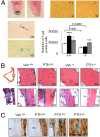
Altered PTH-stimulated β-catenin signaling in Postn−/− mice. (A, Left) Pellet of calvaria osteoblasts shows X-Gal staining in the Postn+/+ but not Postn−/−; (Right) X-Gal staining of calvaria osteoblasts after 14 d in culture. Nuclear and cytoplasm staining are indicated by an arrow and asterisk, respectively. The histogram shows the effects of PTH and Postn on the numbers of X-Gal+ cells. *P < 0.05, **P < 0.01, ***P < 0.001, ****P < 0.0001 vs. vehicle. Bars show means (± SEM). Open bars, vehicle; closed bars, PTH 10−7 M; shaded bars, recombinant periostin (2 μg/mL). (B) X-Gal staining of midshaft tibia from TOPGAL;Postn+/+ and TOPGAL;Postn−/− mice. (C) Immunohistochemical staining of periostin protein expression (brown) and X-Gal (blue). Ec, endocortical; Ps, Periosteum surfaces.
Fig. 5.
Fig. 5.
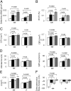
Effects of Sclerostin blocking antibodies (Sost-Ab) on bone response to iPTH. (A) Femur BMD gain. (B) BV/TV of the distal femur. (C) CtTV and CtBV of the midshaft femur. (D) Biomechanical properties of the cortical tibia. (E) Moment of inertia. (F) Difference of seric osteocalcin levels between the end and the beginning of the treatments. *P < 0.05, **P < 0.01, ***P < 0.001, ****P < 0.0001 PTH vs. vehicle in pretreatment (Sost-Ab or control-Ab) and genotype (Postn+/+ or Postn−/−). Bars shows mean (± SEM). Open bars, Control-Ab+vehicle; closed bars, Control-Ab+PTH (40 μg⋅kg⋅d); light shaded bars, Sost-Ab+vehicle; dark shaded bars, Sost-Ab+PTH (40 μg⋅kg⋅d).
Fig. 6.
Fig. 6.
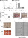
Proliferation and differentiation of Postn-deficient osteoblast. (A) Proliferation was evaluated by thymidine 3H in Postn+/+ and Postn−/− mouse calvaria osteoblasts after 2, 4, and 6 d of culture. **P < 0.01, ***P < 0.001 by unpaired t test compared with Postn+/+ mice. (B) Cell proliferation was evaluated after 6 h of PTH treatment (10−7 M) in Postn+/+ and Postn−/− osteoblasts cultures from calvaria. *P < 0.05 unpaired t test compared with vehicle. (C) ALP production and staining in osteoblasts cultures from Postn+/+ and Postn−/− calvaria mice after 14 d. ***P < 0.001 unpaired t test compared with Postn+/+ mice. (D) Mineralization of Postn+/+ and Postn−/− calvaria evaluated by Alizarin red after 21 and 28 d of culture. (E) Scratch-wound test to assess migration of osteoblast. Bars shows mean (± SEM). Open bars, vehicle Postn+/+; hatch open bars, PTH Postn+/+; shaded bars, vehicle Postn−/−; hatch shaded bars, PTH Postn−/−.
Fig. 7.
Fig. 7.
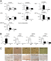
Influence of Postn on PTH-regulated osteoblast/osteocyte gene expression. (A) The relative expression of selected osteoblastic (A) and osteocytic (B) genes was determined by quantitative RT-PCR adjusted to Gapdh after 14 and 28 d of culture. (C) Immunohistochemical analysis of apoptotic cells in response to PTH in longitudinal sections of the proximal tibia and in Postn+/+ and Postn−/− calvaria mice after 14 d of culture. White arrow, caspase 3+ cell; black arrow, caspase 3 cell. (Magnification: Upper, 10×; Lower, 20×.) *P < 0.05, **P < 0.01, ***P < 0.001, ****P < 0.0001 vs. vehicle. Bars shows mean (± SEM). Open bars, vehicle; closed bars, PTH 10−7 M; shaded bars.

References

    1. Kraenzlin ME, Meier C. Parathyroid hormone analogues in the treatment of osteoporosis. Nat Rev Endocrinol. 2011;7:647–656. - PubMed
    1. Kousteni S, Bilezikian JP. The cell biology of parathyroid hormone in osteoblasts. Curr Osteoporos Rep. 2008;6:72–76. - VSports - PubMed
    1. Jilka RL. Molecular and cellular mechanisms of the anabolic effect of intermittent PTH. Bone. 2007;40:1434–1446. - PMC - PubMed
    1. Huang JC, et al. PTH differentially regulates expression of RANKL and OPG. J Bone Miner Res. 2004;19:235–244. - VSports在线直播 - PubMed
    1. Martin TJ, Sims NA. Osteoclast-derived activity in the coupling of bone formation to resorption. Trends Mol Med. 2005;11:76–81. - PubMed

Publication types

MeSH terms

Substances

"V体育2025版" LinkOut - more resources