Skip to main page content
U.S. flag

An official website of the United States government

Dot gov

The . gov means it’s official. Federal government websites often end in . gov or . mil VSports app下载. Before sharing sensitive information, make sure you’re on a federal government site. .

Https

The site is secure. The https:// ensures that you are connecting to the official website and that any information you provide is encrypted and transmitted securely V体育官网. .

. 2013 Jul 11;32(28):3381-9.
doi: 10.1038/onc.2012.342. Epub 2012 Aug 6.

"VSports手机版" Snail1 controls TGF-β responsiveness and differentiation of mesenchymal stem cells

Affiliations

Snail1 controls TGF-β responsiveness and differentiation of mesenchymal stem cells

R Batlle et al. Oncogene. .

Abstract

The Snail1 transcriptional repressor plays a key role in triggering epithelial-to-mesenchymal transition. Although Snail1 is widely expressed in early development, in adult animals it is limited to a subset of mesenchymal cells where it has a largely unknown function. Using a mouse model with inducible depletion of Snail1, here we demonstrate that Snail1 is required to maintain mesenchymal stem cells (MSCs). This effect is associated to the responsiveness to transforming growth factor (TGF)-β1 that shows a strong Snail1 dependence. Snail1 depletion in conditional knockout adult animals causes a significant decrease in the number of bone marrow-derived MSCs. In culture, Snail1-deficient MSCs prematurely differentiate to osteoblasts or adipocytes and, in contrast to controls, are resistant to the TGF-β1-induced differentiation block VSports手机版. These results demonstrate a new role for Snail1 in TGF-β response and MSC maintenance. .


Figures

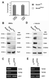
Fig 1
Fig 1. Snail1 induces the expression of markers of activated fibroblasts
Cell populations were transfected with either pcDNA3-Snail1-HA or pcDNA3 control, or with shRNAs specific for Snail1 or a scrambled control. Where mentioned, cell lines were treated with TGF-β1 (5 ng/ml) for 24 hours or for the time indicated. Protein (A, B, D and E, lower panel) or RNA (C, and E upper panel) levels were determined by western blot o semi-quantitative RT-PCR, respectively. Pyruvate Kinase (PyrK) was used as loading control in the western blots; HPRT, in the RT-PCR analysis. The results are representative of three experiments.
Fig 2
Fig 2. Snail1 prevents the differentiation of 3T3-L1 pre-adipocytes
Control cells or 3T3-L1 transfected with control plasmid, Snail1 wild-type or P2A mutant were cultured in regular DME medium or in differentiation medium for two days after the cells arrived to confluence. Culture medium was replaced by DME medium plus 10% FBS (see Methods) and after two more days cells were either stained with Oil Red (panel A) or homogenized in order to get protein cell extracts (panels B and C). Protein expression was determined using antibodies against Snail1, C/EBPα, Glut4, Glucocorticoid receptor (GR) or PyrK as loading control. When indicated, TGF- β1 (5 ng/ml) was supplemented to the medium at the same moment that the cells arrived to confluence and two days later. The figure shows representative results of at least three experiments performed.
Fig 3
Fig 3. Snail1 blocks MSCs differentiation to osteoblasts or adipocytes
Murine MSCs were transfected with pcDNA3-Snail1-HA or the empty plasmid as control. Protein expression was determined by western blot with the indicated antibodies from extracts prepared from MSCs differentiated to osteoblasts (for 10 days) or adipocytes (14 days) as indicated in Methods (A), or not-differentiated (B top, and C). As control for differentiated cells in panel A (not differentiated), cells were incubated with DMEM plus FBS rather than differentiation medium. RNA was also extracted from MSCs transfected or not with Snail1-HA before being challenged to differentiate and analyzed by RT-PCR for TGFB1 and TGFB2 expression (B, bottom). In panel C, TGF-β1 (5 ng/ml) was added to the cell medium 24 hours before preparing the extracts. (D-G); cells were differentiated to osteoblasts (D and E) or adipocytes (F and G); to determine osteoblast differentiation, cells were stained with Alizarin Red (D); alternatively, cell extracts were prepared and AP activity was determined (E, left) or Osx RNA analyzed (E, right). Adipocytes were stained with Oil Red-O (F); the presence of the differentiation marker Glut4 was determined by western blot (G).
Fig 4
Fig 4. Akt is activated by Snail1 ectopic expression in MSCs
(A); the expression of the indicated proteins was determined in undifferentiated MSCs treated with LY (25 μM) for 24 hours before preparing the extracts. As in Figs 3A and B, cells were pretreated for 15 hours in DMEM medium plus 2% FBS. (B and C); Snail1-transfected MSCs were incubated in osteoblasts differentiation medium for 10 days in the presence of LY, MK, or Wortmannin from one day before adding the medium until two days later (see also Fig 7). Differentiation was determined staining with Alizarin Red (B) or measuring AP activity (C). Results are representative, or show the average ± range of three experiments. (D, E); a constitutively active form of Akt (CA-Akt), tagged with an HA-epitope, was transfected to MSCs. Cells were selected, challenged to differentiate for ten days and stained with Alizarin Red (D). The expression of CA-Akt was checked by western blot (E); since Thr308 and Ser473 are mutated in this form (36), CA-Akt activity was determined analyzing S6 phosphorylation.
Fig 5
Fig 5. Snail1 controls the number of bone marrow MSCs in vivo
Femur marrow cells were isolated from Snail1Flox/+ or Snail1Flox/− animals one week after injection of tamoxifen. (A); cells were analyzed by FACS analysis with antibodies against CD45 and CD105 or CD45 and CD90 antibodies. The percentage of CD45-/CD105+ or CD44-/CD90+ cells is presented for both types of animals. The results show the average ± SD of three experiments performed (B and C) Cells were attached to the cell culture plates and grown for other seven days. Protein extracts (panel B) or total RNA (panel C) was prepared and analyzed. In parallel experiments, bone marrow MSCs were obtained from FloxSnail1/del animals prior to treatment with tamoxifen (TAM); cells were then incubated with hidroxi-tamoxifen (TAM-OH) (1 μM) for two days. Twenty four hours later, the indicated proteins (D) or RNAs (E) were analyzed as above.
Fig 6
Fig 6. Snail1 depletion promotes the premature differentiation of MSCs and inhibits the response to TGF-β1
MSCs were obtained from Snail1Flox/− animals prior to treatment with tamoxifen, grown to confluency and incubated with TAM-OH for two days. TAM-OH was removed and, after 24 hours, cells were supplemented with the osteoblasts differentiation medium (A, B and E). The extent of differentiation was determined staining with Alizarin Red (A, E) or measuring AP (B). When indicated cells were treated with TGF-β1 (5 ng/ml) one day before adding TAM-OH as indicated in Methods. (C, D); similar experiments were carried out but differentiating cells to adipocytes as described in Fig 3. Differentiation was determined by Oil Red-O staining (C) four days after initiating the process (note that the result presented in Fig 3 was obtained after 14 days) or analyzing Glut4 expression. (F-H); two days after removal of TAM-OH cells were treated with TGF-β1 for 24 hours; the levels of the indicated RNAs (F and H, lower panel) or proteins (G and H, upper panel) were determined as previously described. (I); chromatin immunoprecipitation (ChIP) assays were performed in control or Snail1-depleted MSCs treated or not with TGF-β as indicated in Methods, to determine Snail1 binding to PTEN promoter. The results show the average ± SD of three experiments performed.
Fig 7
Fig 7. PI3K/Akt inhibitors prevent TGF-β effects on MSC markers and osteogenic differentiation
Panel A; control MSCs were pretreated with DMEM plus 2% FBS for 15 hours and incubated with TGF-β1 (5 ng/ml) or TGF-β plus LY-294002 (25 μM) for 20 hours. Cells extracts were prepared and analyzed by western blot. Panels B, C and D; MSCs were induced to differentiate to osteoblasts; one day before adding the differentiation medium (D-1) TGF-β1 was supplemented with LY or MK when indicated and maintained until D+2. Differentiation was assessed by Alizarin Red Staining (B, D) or measuring AP activity (C) at D+10. Alternatively, TGF-β1 was added at D+1 and maintained until D+8, or LY added at D+4. Panel E; MSCs, one day before reaching confluence and adding the differentiation medium (D-1) or one day after (D+1), were incubated with TGF-β for 20 hours. Cell extracts were prepared an analysed by western blot.

"V体育官网入口" References

    1. Peinado H, Olmeda D, Cano A. Snail, ZEB and bHLH factors in tumour progression: and alliance against the epithelial phenotype? Nat. Rev. Cancer. 2007;7:415–428. - PubMed
    1. Garcia de Herreros A, Peiro S, Nassour M, Savagner P. Snail family regulation and epithelial-mesenchymal transitions in breast cancer progression. J. Mammary Gland Biol Neoplasia. 2010;15:135–147. - PMC - PubMed
    1. Batlle E, Sancho E, Francí C, Domínguez D, Monfar M, Baulida J, et al. The transcription factor snail is a repressor of E-cadherin gene expression in epithelial tumour cells. Nat. Cell Biol. 2000;2:84–89. - PubMed
    1. Cano A, Pérez-Moreno MA, Rodrigo I, Locascio A, Blanco MJ, del Barrio MG, et al. The transcription factor snail controls epithelial-mesenchymal transitions by repressing E-cadherin expression. Nat. Cell Biol. 2000;2:76–83. - PubMed
    1. Solanas G, Porta-de-la-Riva M, Agustí C, Casagolda D, Sánchez-Aguilera F, Larriba MJ, et al. E-cadherin controls beta-catenin and NF-kappaB transcriptional activity in mesenchymal gene expression. J. Cell Sci. 2008;121:2224–2234. - PubMed

Publication types

MeSH terms