Skip to main page content
U.S. flag

An official website of the United States government

Dot gov

The . gov means it’s official. Federal government websites often end in . gov or . mil VSports app下载. Before sharing sensitive information, make sure you’re on a federal government site. .

Https

The site is secure. The https:// ensures that you are connecting to the official website and that any information you provide is encrypted and transmitted securely. V体育官网.

. 2013 Feb;5(1):14-26.
doi: 10.1093/jmcb/mjs042. Epub 2012 Jul 24.

New therapy targeting differential androgen receptor signaling in prostate cancer stem/progenitor vs. non-stem/progenitor cells

Affiliations

VSports注册入口 - New therapy targeting differential androgen receptor signaling in prostate cancer stem/progenitor vs. non-stem/progenitor cells

"VSports手机版" Soo Ok Lee et al. J Mol Cell Biol. 2013 Feb.

Erratum in

Abstract

The androgen deprivation therapy (ADT) to systematically suppress/reduce androgens binding to the androgen receptor (AR) has been the standard therapy for prostate cancer (PCa); yet, most of ADT eventually fails leading to the recurrence of castration resistant PCa. Here, we found that the PCa patients who received ADT had increased PCa stem/progenitor cell population VSports手机版. The addition of the anti-androgen, Casodex, or AR-siRNA in various PCa cells led to increased stem/progenitor cells, whereas, in contrast, the addition of functional AR led to decreased stem/progenitor cell population but increased non-stem/progenitor cell population, suggesting that AR functions differentially in PCa stem/progenitor vs. non-stem/progenitor cells. Therefore, the current ADT might result in an undesired expansion of PCa stem/progenitor cell population, which explains why this therapy fails. Using various human PCa cell lines and three different mouse models, we concluded that targeting PCa non-stem/progenitor cells with AR degradation enhancer ASC-J9 and targeting PCa stem/progenitor cells with 5-azathioprine and γ-tocotrienol resulted in a significant suppression of the tumors at the castration resistant stage. This suggests that a combinational therapy that simultaneously targets both stem/progenitor and non-stem/progenitor cells will lead to better therapeutic efficacy and may become a new therapy to battle the PCa before and after castration resistant stages. .


Figures

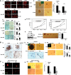
Figure 1
Figure 1
Stem/progenitor cells increase after castration/ADT. (A) Cell line studies. (a) Flow cytometric analysis of CD133+ cells after 1 (red), 3 (yellow), and 5 (green) weeks of 1 µM Casodex® treatment of LNCaP cells. (b) Western blot analysis of CD133 expression after 5 weeks of Casodex® treatment. (B) Mice tumor tissue studies. Immunohistostaining (IHC) results of tumor tissues. (a) Tumors of LNCaP-xenografted mice and (b) tumor tissues of C4-2-xenografted mice. Tumor tissues were obtained before and 10, 20, and 30 days (indicated as B, 10, 20, and 30) after castration and stained with antibodies of CD133, integrin, CK5, and CK8. Quantitation is shown below the staining data (magnification, ×100; inset, ×400). (C) Human tumor tissues studies. IHC of tumor tissues with antibodies of CD133, CD44, CK5, integrin, and CK8. Human tissues were obtained from Tohoku University Hospital, Sendai, Japan, and Chang Gung Memorial Hospital, Linkou, from the same patients, before and after ADT (magnification, ×100; inset, ×400). **P< 0.01.
Figure 2
Figure 2
All PCa cells/tumor tissues contain stem/progenitor and non-stem/progenitor cells. (In figure labels, stem/progenitor and non-stem/progenitor cells were abbreviated as S/P and non-S/P cells, respectively.) (A) Cell line data (LNCaP). (a) Separation of stem/progenitor (1.1%) and non-stem/progenitor (98.9%) cells of the LNCaP cell line by flow cytometry. Antibodies of CD133 and integrin were used. (b) Morphology of stem/progenitor cells in culture. (c) IF staining results of stem/progenitor and non-stem/progenitor cells of the LNCaP cell line using antibodies of markers indicated. The stem/progenitor cells stained positive for CK5, CD133, and integrin, whereas the non-stem/progenitor cells were positive for CK8 and AR (magnification, ×400; inset, 4-b-diamini-2-phenylindole (DAPI) staining). (d) qPCR analysis results showing mRNA expression levels of markers indicated. (e) Western blot analysis of CD133, integrin, and CK8 expressions using cell extracts of parental, stem/progenitor, and non-stem/progenitor cells. Glyceraldehyde-3-phosphate dehydrogenase (GAPDH) antibody was used as a control. (f) The soft agarose colony formation assay results using stem/progenitor and non-stem/progenitor cells of LNCaP cells. (g) Results of xenograft mice study. LNCaP cells, stem/progenitor and non-stem/progenitor cells (1 × 10⁵/site), were injected orthotopically into AP lobes of nude mice. Mice were sacrificed 4 weeks after and the size of the tumor was compared. (h) Invasion and migration assays of stem/progenitor and non-stem/progenitor cells at 1 and 10 nM DHT concentrations. (i) qPCR analysis of AR mRNA expression. (j) Western blot analysis of AR expression using the same cell extracts used in (e). (k) qPCR analysis results of AR mRNA expression when cells were grown in normal culture condition or as spheres on Matrigel coated plates. (B) Tumor tissue data. Tumor tissues were obtained, digested with collagenase (0.28%), DNase I, 10% FCS, one antibiotic/antimycotic, in RPMI. The dissociated cells were further digested with trypsin/EDTA (0.25%) and filtered through strainer. (a) Flow cytometric separation of stem/progenitor and non-stem/progenitor cells obtained from TRAMP tumor tissues (B6 background, 26 weeks old mice). Antibodies of sca-1 and integrin were used in the separation. (b) qPCR analysis result of AR mRNA expression in stem/progenitor and non-stem/progenitor cells isolated from TRAMP mice tumor tissues. (c) IF staining showing marker expressions in stem/progenitor and non-stem/progenitor cells isolated from TRAMP mice tumor tissues. The stem/progenitor cells stained positive for sca-1, CD44, and integrin, whereas the non-stem/progenitor cells were positive for CK8 (magnification, ×400; inset, DAPI staining). (d) IF staining results demonstrating marker expressions in stem/progenitor and non-stem/progenitor cells obtained from C4-2 orthotopic xenografted tumor tissues. The stem/progenitor cells stained positive for CD133 and CK5, whereas the non-stem/progenitor cells were positive for CK8 and AR (magnification, ×400; inset, DAPI staining). (e) Separation of stem/progenitor and non-stem/progenitor cells from human patient tumor tissues (age 51, prostatic adenocarcinoma, Gleason score 6) by collagenase/trypsin digestion as described earlier. (f) IF staining results of marker expressions in stem/progenitor and non-stem/progenitor cells of human patient tumor tissues. The stem/progenitor cells stained positive for CD133, CK5, and integrin and slightly positive for CK8, whereas the non-stem/progenitor cells were strongly positive for CK8 and AR (magnification, ×400; inset, DAPI staining). All experiments were done three times.
Figure 3
Figure 3
Opposite roles of AR in self-renewal/proliferation of stem/progenitor and non-stem/progenitor cells. (In figure labels, stem/progenitor and non-stem/progenitor cells were abbreviated as S/P and non-S/P cells, respectively.) All assays were performed after infection of stem/progenitor and non-stem/progenitor cells with lentivirus carrying vector, AR cDNA, or AR-siRNA. (A) Cell line studies. (a) IF staining result using Ki67 antibody (magnification, ×400). (b) The sphere formation assay. Quantitation on right. (c) The soft agar colony formation assay. Quantitation on right. (d) The MTT assay result at 1 and 10 nM DHT concentrations. (e) Left panel shows AR expression status of injected cells (105) in LNCaP xenograft mice study. Right panel represents tumors obtained from mice of two groups, inoculated with the vector expressing CD133+ or AR expressing CD133+ cells. All experiments were done three times. *P< 0.05 and **P< 0.01. (B) Mice tumor tissue data. (a) BrdU-labeling data (IHC) of stem/progenitor and non-stem/progenitor cells obtained from TRAMP mice tissues (magnification, ×400). (b) Upper two figures represent the sphere formation assay using stem/progenitor cells and lower figures are the results of BrdU-labeling experiment using non-stem/progenitor cells obtained from C4-2 xenograft tumor tissues. (c) The sphere formation assay of stem/progenitor cells obtained from TRAMP mice tissues. Quantitation on right. *P< 0.05 and **P< 0.01. (C) Human tumor tissue data. (a) Ki67 staining (IF staining) using stem/progenitor and non-stem/progenitor cells obtained from human tumor tissues (magnification, ×400; inset, DAPI staining). (b) The sphere formation assay using stem/progenitor cells of human tumor tissues. Quantitation on right.
Figure 4
Figure 4
The in vitro test of targeting stem/progenitor cells by inhibiting activated signaling molecules. (In figure labels, stem/progenitor and non-stem/progenitor cells were abbreviated as S/P and non-S/P cells, respectively.) (A) Western blot analysis using cell extracts of parental, stem/progenitor, and non-stem/progenitor cells of the LNCaP cell line and antibodies indicated. (B) Western blot analysis using cell extracts of stem/progenitor cells after vector (V)/AR carrying lentiviral infection. MTT analysis result of stem/progenitor cells upon γ-TT treatment is with LNCaP (C) and with C4-2 cells (D). (E) Western blot analysis of signaling molecules after treatment with γ-TT. (F) ErbB2 mRNA expressions in LNCaP stem/progenitor and non-stem/progenitor cells. (G) Decrease in ErbB2 mRNA expression in LNCaP stem/progenitor cells upon γ-TT treatment. (H) LNCaP non-stem/progenitor cells were treated with different concentrations of 5-AZA and growth was analyzed. (I) The AR mRNA level in LNCaP-CD133+ cells was analyzed by qPCR after treatment with 5-AZA.
Figure 5
Figure 5
Combined use of γ-TT and 5-AZA acted effectively to kill stem/progenitor cells. (In figure labels, stem/progenitor and non-stem/progenitor cells were abbreviated as S/P and non-S/P cells, respectively.) (A and B) Cell growth analysis using three drugs, γ-TT, and 5-AZA, either individually or two drugs combined. Cells (A with LNCaP stem/progenitor cells and B with C4-2 stem/progenitor cells) were treated with indicated concentrations of drugs for 4 days and cell numbers were counted at the end of incubation. (C) Effect of combined use of drugs on sphere formation. Quantitation is shown in lower panel. (D) Effect of combined use of drugs on colony formation on soft agarose. Quantitation is shown in lower panel. (E) The sphere formation assay using LNCaP stem/progenitor cells after treatment with either Casodex or Drug treatment. (F) The cell growth assay of LNCaP non-stem/progenitor cells with various concentrations of ASC-J9®.
Figure 6
Figure 6
Targeting PCa stem/progenitor and non-stem/progenitor simultaneously blocks the growth of castration resistant tumors. (In figure labels, stem/progenitor and non-stem/progenitor cells were abbreviated as S/P and non-S/P cells, respectively.) (A) Tumors obtained from xenograft mice study. LNCaP cells were orthotopically inoculated into AP lobes of 8 weeks old nude mice. When tumors developed to palpable sizes, mice were castrated and we waited for 2 weeks for tumors to re-develop. Drug treatment started in three groups of mice as indicated (every other day, for 2 weeks). Doses of 0.25 mg/kg 5-AZA and 6.25 mg/kg γ-TT were used. (B) IHC staining of tumor tissues obtained at sacrifice (magnification, ×100; inset, ×400). Quantitation is shown at right. (C) C4-2 cells were orthotopically implanted into AP lobes of the castrated nude mice (8 weeks). When tumors developed, the mice were treated with the combination of ASC-J9® and drugs (γ-TT and 5-AZA) every other day for 3 weeks. Doses of 0.25 mg/kg 5-AZA, 6.25 mg/kg γ-TT, and 50 mg/kg of ASC-J9® were used. (D) IHC staining of tumor tissues obtained at sacrifice (magnification, ×100; inset, ×400). Quantitation at right. (E) C81 cells orthotopically implanted into AP lobes of the castrated nude mice (8 weeks). When tumors developed, the mice were treated with the combination of ASC-J9® and drugs (γ-TT and 5-AZA; same doses as in C4-2 xenograft, C) every other day for 3 weeks. Quantitation at right. (F) IHC staining of tumor tissues obtained at sacrifice (magnification, ×100; inset, ×400).

References

    1. Bisson I., Prowse D.M. WNT signaling regulates self-renewal and differentiation of prostate cancer cells with stem cell characteristics. Cell Res. 2009;19:683–697. - PubMed
    1. Cabrespine A., Guy L., Chollet P., et al. Molecular mechanisms involved in hormone resistance of prostate cancer. Bull. Cancer. 2004;91:747–757. - PubMed
    1. Collins A.T., Habib F.K., Maitland N.J., et al. Identification and isolation of human prostate epithelial stem cells based on alpha(2)beta(1)-integrin expression. J. Cell Sci. 2001;114:3865–3872. - PubMed
    1. Comitato R., Nesaretnam K., Leoni G., et al. A novel mechanism of natural vitamin E tocotrienol activity: involvement of ERbeta signal transduction. Am. J. Physiol. Endocrinol. Metab. 2009;297:E427–E437. - "V体育安卓版" PubMed
    1. Crocoll A., Zhu C.C., Cato A.C., et al. Expression of androgen receptor mRNA during mouse embryogenesis. Mech. Dev. 1998;72:175–178. - PubMed (VSports app下载)

Publication types (VSports手机版)

MeSH terms