Skip to main page content
U.S. flag

An official website of the United States government

Dot gov

The . gov means it’s official. Federal government websites often end in . gov or . mil VSports app下载. Before sharing sensitive information, make sure you’re on a federal government site. .

Https

The site is secure. The https:// ensures that you are connecting to the official website and that any information you provide is encrypted and transmitted securely. V体育官网.

. 2012 Jul 24;5(234):ra52.
doi: 10.1126/scisignal.2002918.

p53 functions in endothelial cells to prevent radiation-induced myocardial injury in mice

Affiliations

"V体育安卓版" p53 functions in endothelial cells to prevent radiation-induced myocardial injury in mice

Chang-Lung Lee et al. Sci Signal. .

Abstract

Radiation therapy, which is used for the treatment of some cancers, can cause delayed heart damage. In the heart, p53 influences myocardial injury that occurs after multiple types of stress. Here, we demonstrated that p53 functioned in endothelial cells to protect mice from myocardial injury after whole-heart irradiation. Mice with an endothelial cell-specific deletion of p53 succumbed to heart failure after whole-heart irradiation as a result of myocardial necrosis, systolic dysfunction, and cardiac hypertrophy. Moreover, the onset of cardiac dysfunction was preceded by alterations in myocardial vascular permeability and density, which resulted in cardiac ischemia and myocardial hypoxia. Mechanistic studies with primary cardiac endothelial cells irradiated in vitro indicated that p53 signaling caused mitotic arrest and protected cardiac endothelial cells from cell death resulting from abnormal mitosis or mitotic catastrophe VSports手机版. Furthermore, mice lacking the cyclin-dependent kinase inhibitor p21, which is a transcriptional target of p53, were also sensitized to myocardial injury after whole-heart irradiation. Together, our results demonstrate that the p53-p21 axis functions to prevent radiation-induced myocardial injury in mice. .


Figures (VSports在线直播)

Fig. 1
Fig. 1
Deletion of p53 by Tie2Cre sensitizes mice to radiation-induced myocardial injury. (A) Representative fluoroscopy image of a mouse treated with whole-heart irradiation. Whole-heart irradiation was performed with a 15 mm circular collimator (red circle) (B and C) Kaplan-Meier survival analysis of Tie2Cre; p53FL/+ and Tie2Cre;p53FL/– mice after a single fraction of 12 Gy or 10 daily fractions of 3 Gy whole-heart irradiation (3 Gy × 10). P value was calculated by long-rank test. (D – G) Histological analysis of the myocardium of moribund Tie2Cre; p53FL/− mice after 12 Gy whole-heart irradiation. Representative sections of the myocardium of a Tie2Cre; p53FL/− mouse 62 days after 12 Gy whole-heart irradiation (D) were stained with hematoxylin and eosin (H&E) (E), anti-sarcomeric α-actin IgG (SαA) (F) or Masson's trichrome (Trichrome) (G). The dashed line in (E), (F) and (G) demarcates the relatively normal appearing myocardium and the region of necrosis (labeled as N). Yellow arrows indicate necrotic cardiomyocytes replaced by blue collagen fibers. (H and I) Evans blue dye uptake (red) in Tie2Cre; p53FL/+ (H) and Tie2Cre; p53FL/− (I) mice 5 weeks after 12 Gy whole-heart irradiation. Cell membranes were counterstained with wheat germ agglutinin (white). Images represent four mice per group. Scale bar, 100 μm.
Fig. 2
Fig. 2
Deletion of p53 by Tie2Cre sensitizes mice to systolic dysfunction and cardiac hypertrophy. (A) Representative echocardiographic recordings from Tie2Cre; p53FL/+ and Tie2Cre; p53FL/− mice prior to and 7 weeks after 12 Gy whole-heart irradiation. (B to E) Changes in fractional shortening (B), heart rate corrected mean velocity of circumferential fiber shortening (mVcfc) (C), left ventricular end-systolic dimension (LVDs) (D) and the left ventricular mass (E) in Tie2Cre; p53FL/+ and Tie2Cre; p53FL/− mice after 12 Gy whole-heart irradiation. Data are presented as mean ± SEM (n=6 mice per group). (F) Ratio of heart weight to body weight (HW/BW) of Tie2Cre; p53FL/+ and Tie2Cre; p53FL/− mice sham irradiated or 7 weeks after 12 Gy whole-heart irradiation. Data are presented as mean ± SEM (n=3 mice per group). (G) Representative images of wheat germ agglutinin (WGA)-stained myocardium of Tie2Cre; p53FL/+ and Tie2Cre;p53FL/− mice sham irradiated or 7 weeks after 12 Gy whole-heart irradiation. Scale bar, 100 μm. (H) Quantification of cardiomyocyte cross-sectional area in WGA-stained myocardium. Data are presented as mean ± SEM (n=3 mice per group). Asterisk shows individual differences (P<0.05) by two-way ANOVA with Bonferroni post-hoc test.
Fig. 3
Fig. 3
Deletion of p53 by Tie2Cre sensitizes mice to vascular injury and chronic ischemia in the myocardium. (A – C) Representative sections of WGA (white) and GS-IB4 (red)-stained myocardium of Tie2Cre; p53FL/+ (A) and Tie2Cre; p53FL/− mice (B and C) 4 weeks after 12 Gy whole-heart irradiation. (D and E) Quantification of GS-IB4 positive myocardial capillaries per high power (200×) field. For Tie2Cre; p53FL/− mice 4 weeks after 12 Gy whole-heart irradiation, vessel density in normal and necrotic regions are scored separately. By 7 weeks, no normal appearing myocardium was present in Tie2Cre; p53FL/− mice. Asterisk shows individual differences (P<0.05) by one-way ANOVA with Bonferroni post-hoc test compared to sham irradiated mice. Data are presented as mean ± SEM (n=3 mice per group). (F) Representative sections of the myocardium of Tie2Cre; p53FL/+; mTmG/+ and Tie2Cre; p53FL/−; mTmG/+ mice 7 weeks after 12 Gy whole-heart irradiation. Myocardial vessels were counterstained with GS-IB4. N marks an area of necrosis. High power magnification image shows disorganized vessels in the area adjacent to a necrotic region. Images represent three mice per group. (G) The hypoxia marker EF5 (red) in Tie2Cre; p53FL/+ and Tie2Cre; p53FL/− mice 4 and 5 weeks after 12 Gy whole-heart irradiation. EF5 positive regions correlated with loss of myocardial capillaries, shown by GS-IB4 staining (white). Images represent three mice per group. Scale bar, 100 μm.
Fig. 4
Fig. 4
Deletion of p53 by VE-Cadherin-Cre sensitizes mice to radiation-induced myocardial injury. (A) Kaplan-Meier survival analysis of VECre; p53FL/+ and VECre;p53FL/− mice after 12 Gy whole-heart irradiation. P value was calculated by long-rank test. (B) A representative hematoxylin and eosin-stained (H&E) section of the heart from VECre; p53FL/− mice 52 days after 12 Gy whole-heart irradiation. Multifocal myocardial necrosis was mainly observed in the epicardium (E) and midmyocardium (M). (C) Evans blue dye (red) uptake in VECre; p53FL/+ and VECre; p53FL/− mice 9 weeks after 12 Gy whole-heart irradiation. Cell membranes were counterstained with WGA (white). Images represent three mice per group. (D) Representative sections of the myocardium of VECre; p53FL/+; mTmG/+ and VECre; p53FL/−; mTmG/+ mice 7 weeks after 12 Gy whole-heart irradiation. Myocardial vessels were counterstained with GS-IB4. An area of necrosis is marked by N. Images represent three mice per group. (E) The hypoxia marker EF5 (red) in VECre; p53FL/+ and VECre; p53FL/− mice 5 and 7 weeks after 12 Gy whole-heart irradiation. EF5 positive regions correlated with loss of myocardial capillaries, shown by GS-IB4 staining (white). Images represent two mice per group. Scale bar, 100 μm.
Fig. 5
Fig. 5
p53 protects cardiac endothelial cells from radiation-induced mitotic catastrophe. Experiments were performed using cardiac endothelial cells from Tie2Cre; p53FL/+ and Tie2Cre; p53FL/− mice at passage 3 to 5. (A – B) Quantification of cell death 4 and 96 hours after various doses of irradiation. Cell survival was determined by propidium iodide exclusion. (C–D) Quantification of the percentage of annexin V positive cells 4 and 72 hours after 12 Gy irradiation. (E and F) Quantification of the percentage of cleaved caspase-3 (Casp3) positive (E) and mitotic ratio (F) of cardiac endothelial cells at various time points after 12 Gy irradiation. (G) Representative images of cells with micronuclei. Cells were stained with anti-γ-H2AX antibody and Hoechst 33324 to label γ-H2AX foci (green) and nuclei (blue), respectively. Arrows indicate cells with the presence of micronuclei. The majority of micronuclei contain unrepaired DNA damage indicated by positive staining for γ-H2AX foci. A representative image of a cell with micronuclei at higher magnification is shown in the inset. (H) Quantification of the percentage of cardiac endothelial cells with micronuclei at various time points after 12 Gy irradiation. Asterisk shows individual differences (P<0.05) by two-way ANOVA with Bonferroni post-hoc test. Data are presented as mean ± SEM (n=3 independent experiments).
Fig. 6
Fig. 6
p21-deficient mice are susceptible to radiation-induced myocardial injury. (A) Kaplan-Meier survival analysis of p21+/− and p21−/− mice after 12 Gy whole-heart irradiation. (B) A representative hematoxylin and eosin-stained (H&E) section of the heart from p21−/− mice 34 days after 12 Gy whole-heart irradiation. Scale bar, 100 μm. (C to E) Changes in fractional shortening (C), heart rate corrected mean velocity of circumferential fiber shortening (mVcfc) (D), and left ventricular mass (E) in p21+/− and p21−/− mice after 12 Gy whole-heart irradiation. Data are presented as mean ± SEM. (F) Ratio of heart weight to body weight (HW/BW) of p21+/− and p21−/− mice 0, 3 and 4 weeks after 12 Gy whole-heart irradiation. (G) Representative images of WGA-stained myocardium in p21+/− and p21−/− mice sham irradiated or 7 weeks after 12 Gy whole-heart irradiation. Scale bar, 100 μm. (H) Quantification of cardiomyocyte cross-sectional area in wheat germ agglutinin-stained myocardium. Data are presented as mean ± SEM (n=3 mice per group). Asterisk shows individual differences (P<0.05) by two-way ANOVA with Bonferroni post-hoc test.
Fig. 7
Fig. 7
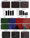
Loss of p21 sensitizes mice to microvascular injury and chronic ischemia in the myocardium. (A) Representative sections of the myocardium stained with Hoechst 33324 (blue) and GS-IB4 (red). Scale bar, 100 μm. (B) Quantification of GS-IB4 positive capillaries per high power (200×) field in the myocardium of p21+/− and p21−/− mice. Asterisk shows individual differences (P<0.05) by two-way ANOVA with Bonferroni post-hoc test. Data are presented as mean ± SEM (n=3 mice for each group). (C) Measurement of vascular permeability in the myocardium of p21+/− and p21−/− mice 3 weeks after 12 Gy whole-heart irradiation. Mice were injected with a mixture of high (2 × 10⁶) and low (1 × 10⁴) molecular weight (molecular weight) dextrans. Blood vessels were stained by anti-CD31 IgG. Images represent three mice per genotype. These results show that dextrans of both low (red) and high (green) MWs are restricted in blood vessels in irradiated p21+/− mice. In contrast, in irradiated p21−/− mice, the high MW dextran is retained in blood vessels, whereas the low MW dextran (red) leaks out of blood vessels in the damaged regions. Images represent three mice per group. (D) The hypoxia marker EF5 (red) in p21+/− and p21−/− mice 3 and 4 weeks after 12 Gy whole-heart irradiation. EF5 positive regions correlated with loss of myocardial capillaries, shown by GS-IB4 staining (white). Images represent two mice per group. Scale bar, 100 μm.

"VSports" References

    1. Gudkov AV, Komarova EA. The role of p53 in determining sensitivity to radiotherapy. Nat Rev Cancer. 2003;3:117. - PubMed
    1. Gudkov AV, Komarova EA. Pathologies associated with the p53 response. Cold Spring Harb Perspect Biol. 2010;2:a001180. - PMC - PubMed
    1. Sano M, et al. p53-induced inhibition of Hif-1 causes cardiac dysfunction during pressure overload. Nature. 2007;446:444. - PubMed
    1. Matsusaka H, et al. Targeted deletion of p53 prevents cardiac rupture after myocardial infarction in mice. Cardiovas Res. 2006;70:457. - PubMed
    1. Leri A, et al. Ablation of telomerase and telomere loss leads to cardiac dilatation and heart failure associated with p53 upregulation. The EMBO journal. 2003;22:131. - PMC - PubMed

VSports - Publication types

"VSports手机版" MeSH terms

V体育ios版 - LinkOut - more resources