Skip to main page content (V体育安卓版)
U.S. flag

An official website of the United States government

Dot gov

The . gov means it’s official. Federal government websites often end in . gov or . mil VSports app下载. Before sharing sensitive information, make sure you’re on a federal government site. .

Https

The site is secure. The https:// ensures that you are connecting to the official website and that any information you provide is encrypted and transmitted securely V体育官网. .

. 2012 Aug 15;72(16):4141-53.
doi: 10.1158/0008-5472.CAN-11-3834. Epub 2012 May 22.

"V体育安卓版" Perturbation of Rb, p53, and Brca1 or Brca2 cooperate in inducing metastatic serous epithelial ovarian cancer

Affiliations

Perturbation of Rb, p53, and Brca1 or Brca2 cooperate in inducing metastatic serous epithelial ovarian cancer (V体育官网)

Ludmila Szabova et al. Cancer Res. .

Abstract

The majority of human high-grade serous epithelial ovarian cancer (SEOC) is characterized by frequent mutations in p53 and alterations in the RB and FOXM1 pathways. A subset of human SEOC harbors a combination of germline and somatic mutations as well as epigenetic dysfunction for BRCA1/2. Using Cre-conditional alleles and intrabursal induction by Cre-expressing adenovirus in genetically engineered mice, we analyzed the roles of pathway perturbations in epithelial ovarian cancer initiation and progression. Inactivation of RB-mediated tumor suppression induced surface epithelial proliferation with progression to stage I carcinoma. Additional biallelic inactivation and/or missense p53 mutation in the presence or absence of Brca1/2 caused progression to stage IV disease. As in human SEOC, mice developed peritoneal carcinomatosis, ascites, and distant metastases. Unbiased gene expression and metabolomic profiling confirmed that Rb, p53, and Brca1/2-triple mutant tumors aligned with human SEOC, and not with other intraperitoneal cancers VSports手机版. Together, our findings provide a novel resource for evaluating disease etiology and biomarkers, therapeutic evaluation, and improved imaging strategies in epithelial ovarian cancer. .


Figures

Figure 1
Figure 1
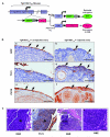
Cre-mediated induction of genetic events in OSE cells by intra-bursal Adeno-Cre injection. A, Scheme depicting the targeting strategy. B, Expression of GFP, T121 antigen and CK18 in serial sections from ovaries of a TgK18GT121tg/+ mouse 3 months pi. GFP expression (a, brown, arrows) and lack of T121 expression (c) in uninduced OSE. Occasional GFP staining (b, arrows) and T121 expression (d, brown, arrows) in the induced OSE. Note that the OSE is hyperplastic with increased numbers of tightly packed columnar cells. IHC staining for CK18 (brown) depicts epithelial cells (e, f, arrows). C, CIS (*) in the induced OSE of a TgK18GT121tg/+ mouse 6 months pi (a). IHC confirming the expression of T121 (b; brown). SEOC with minimal invasion into underlying ovary in a TgK18GT121tg/+ mouse 24 months pi (c). Note unaffected fimbriae and oviduct. Higher magnification of SEOC (d). Scale bars in B represent 50 μm and in C 100 μm.
Figure 2
Figure 2
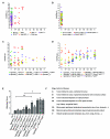
Histopathological contribution of single and compound genetic events in the transformation of OSE. A, RB-TS pathway inactivation in TgK18GT121tg/+ mice is sufficient to initiate surface epithelial proliferation and early SEOC. B, Individual or combined inactivation of Brca1, Brca2 and/or p53 are insufficient to cause ovarian epithelial tumorigenesis. C, Combination of TgK18GT121 and p53 alleles causes SEOC. D, Disease staging in mice with compound induction of RB-TS, p53 and Brca1 or Brca2. N: normal, HP: hyperplasia, CIS: carcinoma in situ. E, Statistical evaluation of disease progression based on histology score (HP=0.25, CIS=0.5, Stage IA=1.0, IB=1.5, IIA=2.0, IIB=2.5, IIIA=3.0, IIIB=3.5, IV=4). Mice removed from study before the age of 3 months were not included in the statistical analysis. Bars depict group mean histology score with error bars representing standard error of the means. One way-ANOVA with Bonferroni’s Multiple Comparison post-test was used to test for significant differences between genotypes. F, Explanation of the histopathological staging system used. The classification was adopted from FIGO used in human ovarian carcinomas.
Figure 3
Figure 3
Perturbation of Rb-TS, p53 and/or Brca1/2 in the OSE induces metastatic serous epithelial ovarian carcinomas. A, MRI of a TgK18GT121tg/+/Brca1fl/fl/p53R172H/fl mouse showing a large ovarian tumor on the induced ovary. B, Abdominal distention and presence of ascites in the same mouse. C, Ovarian tumor and normal non-injected ovary of the same mouse 190 days pi. D, Early SEOC originating from the OSE (50×). Note intact bursa and unaffected oviduct. Range of different histological SEOC features: E, Papillary histology of SEOC shown in D; F, poorly differentiated glandular and microcystic histology; G, micropapillary/filigree; H, papillary and I, solid tumor histology. J, Intraperitoneal spreading (carcinomatosis) on serosal surface of liver (arrow). K, Pleural metastasis (arrow), note alveolar spaces in underlying lung parenchyma (*). L, Liver micrometastases showing the presence of papillary carcinoma within distended hepatic sinusoids (arrow and arrowhead). Scale bar in C represents 1 cm, in D to L 100μm.
Figure 4
Figure 4
Immunophenotype of SEOC arising from the mouse ovarian surface and oviduct epithelium, as well as abdominal carcinomatosis and human peritoneal mesothelioma. Normal mouse OSE (a, g, m s), carcinomas arising from the OSE (b, h, n, t), abdominal carcinomatosis (c, i, o, u), normal oviduct epithelium (d, j, p, v), adenocarcinomas arising from the oviduct (e, k, q, w) and human DMPM (f, l, r, x). Rare expression of CK5 indictated by arrow in w. Scale bars represent 200μm.
Figure 5
Figure 5
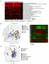
Gene expression patterns and molecular subtypes of mouse SEOC mirror high grade human SEOC. A, Top ranked pathways enriched in tumors versus normals based on SLEPR analysis using Biocarta terms with inclusion of manually curated TCGA pathways (*). The enrichment scores were directly used to create the heatmap in ranked order (higher ranked genes at the top based on the permutated p-values of each pathway). Red, enriched significantly; black, no significant enrichment based on enrichment score of individual sample less than 1.3 (p>0.05) or Gene Hits less than 2. B, PCA of merged microarray data from mouse SEOC and four histological subtypes of human ovarian cancer [GSE6008; (10)]. C, Microarray data from mouse SEOC were merged and clustered together with human SEOC using the TCGA classifier genes (6). Genes in the heatmap were ordered based on their predictive classes of Differentiated, Immunoreactive, Mesenchymal and Proliferative tumor subtypes. The rows depict human (black) or mouse (grey) samples. The data in its original log2 intensity were scaled for purpose of display in heatmap: mean as 0 and standard deviation as 1 for each gene across all samples. D, PCA plot of the top two principal components for mouse tumors and a variety of subtypes of human ovarian carcinomas in the subset of the merged human-mouse array dataset containing the available human-mouse homologous genes out of 189-gene signature previously described to distinguish DMPM from SEOC (11).
Figure 6
Figure 6
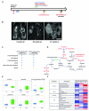
Metabolomic profiling of blood during SEOC progression. A, Experimental design. B, Representative MRI scans from mice 8, 20 and 27 weeks pi, respectively. Arrow indicates the injected ovary, T indicates the ovarian tumor. Scale bar represents 1cm. C, Similar changes in human ovarian cancer tissue (38) and murine whole blood metabolome (this study). D, Integrative model of metabolomic changes observed in the blood of mice with SEOC compared to their littermate controls. Metabolites whose abundance increased are shown in red, decreased in green and unchanged or not detected in black. E, Boxplots comparing the abundance distribution of select metabolites between animals with metastatic ovarian cancer and their littermate (wt) controls at the terminal time points. F, Heat maps depicting temporal metabolite changes in SEOC compared to WT mice. The colors display each group mean level to the median for that compound, such that white represents the median, saturated red represents a two-fold increase relative to and saturated blue is one-half the median value.

References

    1. Jemal A, Siegel R, Xu J, Ward E. Cancer statistics, 2010. CA: a cancer journal for clinicians. 2010;60:277–300. - PubMed
    1. Miller DS, Blessing JA, Krasner CN, Mannel RS, Hanjani P, Pearl ML, et al. Phase II evaluation of pemetrexed in the treatment of recurrent or persistent platinum-resistant ovarian or primary peritoneal carcinoma: a study of the Gynecologic Oncology Group. J Clin Oncol. 2009;27:2686–91. - PMC - PubMed
    1. Jemal A, Murray T, Ward E, Samuels A, Tiwari RC, Ghafoor A, et al. Cancer statistics, 2005. CA: a cancer journal for clinicians. 2005;55:10–30. - PubMed
    1. Neesham D. Ovarian cancer screening. Aust Fam Physician. 2007;36:126–8. - PubMed
    1. Cesario S. Advances in the early detection of ovarian cancer: How to hear the whispers early. Nurs Womens Health. 14:222–34. - PubMed (VSports最新版本)

Publication types

MeSH terms