Skip to main page content (VSports最新版本)
U.S. flag

An official website of the United States government

Dot gov

The . gov means it’s official. Federal government websites often end in . gov or . mil VSports app下载. Before sharing sensitive information, make sure you’re on a federal government site. .

Https

The site is secure. The https:// ensures that you are connecting to the official website and that any information you provide is encrypted and transmitted securely. V体育官网.

. 2012 Jul 20;287(30):25344-52.
doi: 10.1074/jbc.M111.321760. Epub 2012 Apr 30.

Cyclin K-containing kinase complexes maintain self-renewal in murine embryonic stem cells (VSports手机版)

Affiliations

"V体育安卓版" Cyclin K-containing kinase complexes maintain self-renewal in murine embryonic stem cells

Qian Dai et al. J Biol Chem. .

Abstract

Protein phosphorylation plays an important role in the regulation of self-renewal and differentiation of embryonic stem cells. However, the responsible intracellular kinases are not well characterized VSports手机版. Here, we discovered that cyclin K protein was highly expressed in pluripotent embryonic stem cells but low in their differentiated derivatives or tissue-specific stem cells. Upon cell differentiation, the level of cyclin K protein was decreased. Furthermore, knockdown of cyclin K led to cell differentiation, which could be rescued by an expression construct resistant to RNA interference. Surprisingly, cyclin K did not interact with CDK9 protein in cells as thought previously. Instead, it associated with CrkRS (also known as CDK12) and CDC2L5 (also known as CDK13). Similar to cyclin K, both CDK12 and CDK13 proteins were highly expressed in murine embryonic stem cells and were decreased upon cell differentiation. Importantly, knockdown of either kinase resulted in differentiation. Thus, our studies have uncovered two novel protein kinase complexes that maintain self-renewal in embryonic stem cells. .


Figures

FIGURE 1.
FIGURE 1.
Identification of the physiological form of CycK protein. A, expression of CycK protein in different human (293 and HeLa) and murine (mES and F9) cell extracts was analyzed by Western blotting using a commercial anti-CycK antibody (Sigma). 293, HEK 293; mES, mouse embryonic stem cell. B, diagram of two putative human CycK isoforms. The two isoforms are identical in the N-terminal 307 amino acid residues. Epitope peptide (40–56 amino acids) was chemically synthesized and used to generate anti-CycK antibody. C, equal amount of HeLa cell lysate was separated in parallel on the same SDS-PAGE gel and probed separately by crude antiserum (Before) or affinity-purified antibody (After). D, detection of CycK protein in HeLa cell extract by affinity-purified antibody without or with prior neutralization by epitope peptide. E, CycK was probed by Western blotting in extracts from untransfected HeLa cells (−), cells stably transfected with scramble (scra), or two shRNA constructs specific for human CycK (hK-1 and hK-2). Actin was used as a loading control. F, quantification of CycK mRNA by qPCR in cells as indicated in E. The expression of CycK was normalized to actin level and relative to the expression level in cells transfected with scramble shRNA. Expression ratios are average measurements from three independent analyses; S.D. are shown.
FIGURE 2.
FIGURE 2.
CycK protein is highly expressed in mouse ES cells. A, The expression profiles of CycK and CDK9 were analyzed in various murine tissues. Equal amount of protein extract from each tissue, determined by BCA protein assay, was probed by Western blotting. B, CycK expression was analyzed by Western blotting (left) and qPCR (right) in ES and MEF cells. The expression of CycK was normalized to actin level, and expression ratios are average measurements from three independent analyses; S.D. are shown. C, MEF cells growing at indicated confluency were collected and analyzed for CycK by Western blotting. D, MEF cells at ∼30% confluency were deprived of serum for 12 h and then stimulated by indicated concentration of serum for another 12 h before harvest. CycK was analyzed by Western blotting. E, MEF cells were treated with Ultraviolet C (40 J/m2) and recovered in growth medium for indicated time before harvest. F, ES cells were induced to differentiate in vitro for 21 days. Cells were collected at the indicated time and analyzed for CycK expression. G, detection of CycK and Oct4 by indirect immunofluorescence. Background fluorescence was shown on the lower panel when only secondary antibodies (2nd) were used. DNA was revealed by DAPI staining. w/o, without. mus., muscle; pan., pancreas.
FIGURE 3.
FIGURE 3.
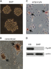
CycK protein is not expressed in dermal stem cells. A, the morphology of dermal stem cells observed under microscope. B, dermal stem cells were induced in vitro to generate osteoblasts (indicated by arrows), revealed by Alizarin Red S staining for calcium. C, dermal stem cells were induced in vitro to generate adipocytes (indicated by arrows), revealed by Oil Red O staining for lipid deposits. D, expression of CycK was analyzed in ES and dermal stem cell extracts by Western blotting.
FIGURE 4.
FIGURE 4.
CycK protein maintains self-renewal in mouse ES cells. A, transient knockdown by scramble (scra), human-specific (hK-1 and hK-2), or murine-specific (mK-1) shRNA constructs in murine ES cells. Knockdown efficiency was analyzed by Western blotting. B, stable knockdown by scramble or mK-1 shRNA. Cell differentiation was indicated by negative AP staining as well as flat cell morphology. C, protein expression was analyzed by Western blotting in extracts from untransfected ES cells (−), cells stably transfected with scramble, or mK-1 shRNA. Cells stably transfected with mK-1 were also trypsinized, reseeded, and grown for 7 days before harvest (mK-1-split). Most cells did not attach upon passage. The remaining cells grew in an ES cell-like morphology. D, rescue of CycK expression by an RNAi-resistant CycK expression vector (CycK-R). Cell extracts from untransfected cells and cells transiently transfected with mK-1, CycK-R, or both were analyzed by Western blotting. E, rescue of self-renewal by CycK-R at the presence of mK-1 shRNA. Cells were stably transfected with indicated constructs and stained by AP. F, stable transfection with another CycK-specific shRNA (mK-2) led to differentiation, revealed by loss of Oct4 immunofluorescence staining. Background fluorescence was shown on the lower panel when only secondary antibody (2nd) was used. G, protein expression was examined by Western blotting in extracts from cells stably transfected with scramble or mK-2 shRNA. DNA was revealed by DAPI staining.
FIGURE 5.
FIGURE 5.
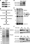
Cyclin K interacts with CDK12 and CDK13 but not CDK9. A, endogenous CDK9 was immunoprecipitated from ES cell extract, followed by Western blotting (WB) with indicated antibodies. B, the experimental flowchart to identify CycK-associated kinases by an unbiased proteomic approach. C, identical purification procedure was carried out using extracts from naïve NIH 3T3 cells (Mock) or cells stably expressing tagged CycK (FH-CycK). Purified materials were visualized by silver. Specific polypeptides in FH-CycK were analyzed by LC-MS/MS. D, purified materials in C were analyzed by Western blotting with indicated antibodies. E, identical purification procedure was carried out using extracts from naïve HEK293 cells (Mock) or two cell lines stably expressing FLAG-tagged CDK12 (CDK12-F). Purified materials were visualized by silver. Specific polypeptides in CDK12-F were analyzed by LC-MS/MS. Identity of CycK was revealed by Western blotting. F, domain mapping of CDK12 and CDK13. cDNAs encoding individual domains were transfected into HEK293 cells, followed by anti-FLAG immunoprecipitation, and a sequential Western blotting with anti-CycK as well as FLAG antibodies. In, Input.
FIGURE 6.
FIGURE 6.
Knockdown of CDK12 or CDK13 leads to ES cell differentiation. A, differentiation of ES cells in vitro. Cells were induced to differentiate by withdrawing LIF and growing in suspension for 7 days (7d), followed by adherent growth for another 7 days (14d). B, protein expression was analyzed by Western blotting in ES cells and differentiated cells as described in A. C, protein expression was analyzed in cells stably transfected with scramble, CDK12-, or CDK13-specific shRNA. D, Oct4 staining was lost in cells stably transfected with CDK12- or CDK13-specific shRNA but not scramble shRNA. Background fluorescence was shown on the lower panel when only secondary antibody (2nd) was used. DNA was revealed by DAPI staining. E, expression levels of pluripotency genes were analyzed by qPCR in cells stably transfected with scramble (scra), CycK, CDK12, or CDK13-specific shRNA. Expression levels were normalized to actin level and relative to the expression levels of the same transcripts in cells transfected with scramble shRNA. Expression ratios are average measurements from three independent analyses; S.D. are shown. F, expression levels of marker genes representative of differentiation were analyzed by RT-PCR in cells stably transfected with scramble, CycK-, CDK12-, or CDK13-specific shRNA. Fgf5 and Sox9 (ectoderm), T (mesoderm), Pdx1, Gata4 and Sox17 (endoderm), Hand1 and Cdx2 (trophectoderm), Pax3 (myogenesis), Slug (EMT), and Dermo (dermal differentiation).

References

    1. Evans M. (2011) Discovering pluripotency: 30 years of mouse embryonic stem cells. Nat. Rev. 12, 680–686 - PubMed
    1. Yu J., Thomson J. A. (2008) Pluripotent stem cell lines. Genes Dev. 22, 1987–1997 - PMC - PubMed
    1. Young R. A. (2011) Control of the embryonic stem cell state. Cell 144, 940–954 - PMC (VSports app下载) - PubMed
    1. Jaenisch R., Young R. (2008) Stem cells, the molecular circuitry of pluripotency and nuclear reprogramming. Cell 132, 567–582 - PMC - PubMed
    1. Saxe J. P., Tomilin A., Schöler H. R., Plath K., Huang J. (2009) Post-translational regulation of Oct4 transcriptional activity. PloS One 4, e4467. - PMC (VSports手机版) - PubMed

Publication types

MeSH terms

Substances