"VSports注册入口" Skip to main page content
U.S. flag

An official website of the United States government

Dot gov

The . gov means it’s official VSports app下载. Federal government websites often end in . gov or . mil. Before sharing sensitive information, make sure you’re on a federal government site. .

Https

The site is secure. The https:// ensures that you are connecting to the official website and that any information you provide is encrypted and transmitted securely. V体育官网.

. 2012;7(4):e35128.
doi: 10.1371/journal.pone.0035128. Epub 2012 Apr 13.

Breast cancer cells induce stromal fibroblasts to secrete ADAMTS1 for cancer invasion through an epigenetic change

Affiliations

Breast cancer cells induce stromal fibroblasts to secrete ADAMTS1 for cancer invasion through an epigenetic change

Shiaw-Wei Tyan et al. PLoS One. 2012.

Abstract

Microenvironment plays an important role in cancer development. We have reported that the cancer-associated stromal cells exhibit phenotypic and functional changes compared to stromal cells neighboring to normal tissues. However, the molecular mechanisms as well as the maintenance of these changes remain elusive. Here we showed that through co-culture with breast cancer cells for at least three to four passages, breast normal tissue-associated fibroblasts (NAFs) gained persistent activity for promoting cancer cell invasion, partly via up-regulating ADAM metallopeptidase with thrombospondin type 1 motif, 1 (ADAMTS1) VSports手机版. Furthermore, we demonstrated that the DNA methylation pattern in the ADAMTS1 promoter has no alteration. Instead, the loss of EZH2 binding to the ADAMTS1 promoter and the resulting decrease of promoter-associated histone H3K27 methylation may account for the up-regulation of ADAMTS1. Importantly, the lack of EZH2 binding and the H3K27 methylation on the ADAMTS1 promoter were sustained in cancer cell-precocultured NAFs after removal of cancer cells. These results suggest that cancer cells are capable of inducing stromal fibroblasts to secrete ADAMTS1 persistently for their invasion and the effect is epigenetically inheritable. .


Conflict of interest statement

Competing Interests: Based on UCI policy, WHL declares that he serves as member of board of directors of GeneTex Inc. This arrangement has been reviewed and approved by the COI committee of UCI. This does not alter the authors' adherence to all the PLoS ONE policies on sharing data and materials V体育安卓版.

Figures

Figure 1
Figure 1. Pre-coculture with breast cancer cells enhanced fibroblast's ability to promote cancer cell invasion.
(A) The diagram of the co-culture protocol. NAF 200N.P7 co-cultured with MDA-MB-468 cells for four passages is indicated as 200N.E1-E4, respectively. Each of 200N.E1-E4 was propagated in the absence of MDA-MB-468 cell for passages from P1 to P3. (B) The conditional media derived from CAF 199C.P10 and cancer cell-precocultured NAF 200N.E4.P3 enhanced the invasion ability of MDA-MB-468 cells and MDA-MB-231 cells. Data are shown as mean ± SD from triplicate experiments. Statistical significance was evaluated by Student's t-test. * P<0.05.
Figure 2
Figure 2. Pre-coculture with breast cancer cells increased ADAMTS1 mRNA levels in NAFs.
(A–C) Quantitative real-time RT-PCR analysis revealed that ADAMTS1 mRNA levels were higher in CAFs than in the corresponding NAFs in ten breast cancer patients (A), and that ADAMTS1 mRNA levels in CAF 199C.P10 and NAF 200N.E4.P3 were higher than in NAF 200N.P10 and 200N.E1-E3.P3 (B). Similar results are shown using the pairs of CAF 244C/NAF 245N and CAF 259C/NAF 260N (C). (D) The ADAMTS1 protein level in NAF 200N.E4.P3, but not NAF 200N.E1-E3.P3, was enhanced to the similar level in CAF 199C.P10, compared to NAF 200N.P10. (E) ELISA indicated that the ADAMTS1 protein level in cultured medium derived from NAF 200N.E4.P3 was higher than NAF 200N.E1-E3.P3 and NAF 200N.P10. (F) ADAMTS1 mRNA level in SK-BR-3 cell-precocultured NAF 200N.S4.P3 was also higher than in NAF 200N.P10 and 200N.S1-S3.P3. Data are shown as mean ± SD from triplicate experiments. Statistical significance was evaluated by Student's t-test. * P<0.05.
Figure 3
Figure 3. ADAMTS1 secreted from stromal fibroblasts enhanced cancer cell invasion.
(A) Sequestration of the ADAMTS1 activity in the conditional medium derived from CAF 199C.P10 using an anti-ADAMTS1 antibody reduced invasion ability of MDA-MB-468 cells (left) and MDA-MB-231 cells (right) in an anti-ADAMTS1 Ab dose-dependent manner. (B) Neutralization of the ADAMTS1 activity in the conditional medium derived from NAF 200N.E4.P3 using an anti-ADAMTS1 antibody also reduced invasion ability of MDA-MB-468 cells (left) and MDA-MB-231 cells (right). (C) Western shows the efficient inhibition of ADAMTS1 protein level by three independent shRNAs. (D) The conditional medium derived from CAF 199C.P10 depleted of ADAMTS1 by shRNAs exhibited significantly lower activity for invasion of MDA-MB-468 cells (left) and MDA.MB-231 cells (right). (E) The conditional media derived from NAF 200N.P10 with exogenous ADAMTS1 expression (left, western) enhanced invasion of MDA-MB-468 cells (middle) and MDA-MB-231 cells (right). Data are shown as mean ± SD from triplicate experiments. Statistical significance was evaluated by Student's t-test. * P<0.05.
Figure 4
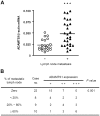
Figure 4. ADAMTS1 expression in CAF correlated with lymph node metastasis.
(A) A scatter dot plot of ADAMTS1 mRNA levels in CAFs assessed using quantitative real-time RT-PCR analysis. The ADAMTS1 mRNA levels in CAFs of patients with lymph node metastasis (n = 27) were significantly higher than in those of patients without lymph node metastasis (n = 22) (P = 0.0003). Data are shown as mean ± SD of triplicate samples. Statistical significance was evaluated by Student's t-test. *** P<0.001. (B) The relationship between ADAMTS1 expression and lymph node metastasis was analyzed using Fisher's exact test. ADAMTS1 expression levels in CAFs derived from patients with ≥60% lymph node metastasis were significantly higher than those of patients with ≤60% lymph node metastasis or with zero lymph node metastasis (P = 0.001). Relative quantity (RQ) of ADAMTS1 expression compared to beta-actin expression: +, 0.001≤RQ≤0.025; ++, 0.025
Figure 5
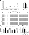
Figure 5. Increase of ADAMTS1 mRNA correlated with reduction of ADAMTS1 promoter-associated H3K27me3 and EZH2 binding through passages.
(A) ADAMTS1 mRNA levels in indicated NAF derivatives were analyzed and shown gradually increased in NAF 200N.E4 from P0 to P3 and maintained from P3 to P5. (B) MeDIP indicates that ADAMTS1 promoter-associated DNA methylation level was similar in NAF 200N.P10, CAF 199C.P10 and NAF 200N.E4.P3 cells. (C) Bisulfite sequencing indicates that ADAMTS1 promoter in CAF 199C.P10, NAF 200N.P10 and NAF 200N.E4.P3 cells was hypomethylated. Closed circle: methylated cytosine. (D) ADAMTS1 promoter-associated H3K27me3 and EZH2 binding was decreased in CAF 199C.P10 and NAF 200N.E4.P3 cells. (B and D) MeDIP (B) or ChIP assays (D) using antibody against H3ace, H3K4me3, H3K9me3, H3K27me3, H3K36me3, H3K79me3 or EZH2 was performed in NAF 200N.P10 (white bars), CAF 199C.P10 (black bars) and NAF 200N.E4.P3 (gray bars) cells, followed by real-time PCR analysis. (E) ADAMTS1 promoter-associated H3K27me3 and EZH2 binding were decreased in NAF 200N.E4.P3 and maintained through passages to NAF 200N.E4.P5. ChIP assays using antibody against H3K27me3 (black bars) or EZH2 (white bars) were performed in cells indicated, followed by real-time PCR analysis. (B, D and E) PCR amplification was performed with DNA primers against the ADAMTS1 promoter region from −431 bp to −217 bp. Data are shown as mean ± SD from triplicate experiments. Statistical significance was evaluated by Student's t-test. * P<0.05. NS: No significant difference.

VSports在线直播 - References

    1. Hu M, Polyak K. Microenvironmental regulation of cancer development. Curr Opin Genet Dev. 2008;18:27–34. - "V体育官网入口" PMC - PubMed
    1. Tlsty TD, Coussens LM. Tumor stroma and regulation of cancer development. Annu Rev Pathol. 2006;1:119–150. - PubMed (V体育平台登录)
    1. Mueller MM, Fusenig NE. Friends or foes - bipolar effects of the tumour stroma in cancer. Nat Rev Cancer. 2004;4:839–849. - PubMed
    1. Tyan SW, Kuo WH, Huang CK, Pan CC, Shew JY, et al. Breast cancer cells induce cancer-associated fibroblasts to secrete hepatocyte growth factor to enhance breast tumorigenesis. PLoS One. 2011;6:e15313. - PMC - PubMed
    1. Allinen M, Beroukhim R, Cai L, Brennan C, Lahti-Domenici J, et al. Molecular characterization of the tumor microenvironment in breast cancer. Cancer Cell. 2004;6:17–32. - "VSports app下载" PubMed

Publication types

MeSH terms