"VSports手机版" Skip to main page content
U.S. flag

An official website of the United States government

Dot gov

The . gov means it’s official. Federal government websites often end in . gov or . mil. Before sharing sensitive information, make sure you’re on a federal government site. VSports app下载.

Https

The site is secure. The https:// ensures that you are connecting to the official website and that any information you provide is encrypted and transmitted securely V体育官网. .

. 2011 Nov;7(11):e1002341.
doi: 10.1371/journal.ppat.1002341. Epub 2011 Nov 3.

"VSports手机版" The critical role of Notch ligand Delta-like 1 in the pathogenesis of influenza A virus (H1N1) infection

Affiliations

The critical role of Notch ligand Delta-like 1 in the pathogenesis of influenza A virus (H1N1) infection

VSports - Toshihiro Ito et al. PLoS Pathog. 2011 Nov.

"VSports在线直播" Abstract

Influenza A viral infections have been identified as the etiologic agents for historic pandemics, and contribute to the annual mortality associated with acute viral pneumonia. While both innate and acquired immunity are important in combating influenza virus infection, the mechanism connecting these arms of the immune system remains unknown. Recent data have indicated that the Notch system is an important bridge between antigen-presenting cells (APCs) and T cell communication circuits and plays a central role in driving the immune system to overcome disease. In the present study, we examine the role of Notch signaling during influenza H1N1 virus infection, focusing on APCs. We demonstrate here that macrophages, but not dendritic cells (DCs), increased Notch ligand Delta-like 1 (Dll1) expression following influenza virus challenge VSports手机版. Dll1 expression on macrophages was dependent on retinoic acid-inducible gene-I (RIG-I) induced type-I IFN pathway, and not on the TLR3-TRIF pathway. We also found that IFNα-Receptor knockout mice failed to induce Dll1 expression on lung macrophages and had enhanced mortality during influenza virus infection. Our results further showed that specific neutralization of Dll1 during influenza virus challenge induced higher mortality, impaired viral clearance, and decreased levels of IFN-γ. In addition, we blocked Notch signaling by using γ-secretase inhibitor (GSI), a Notch signaling inhibitor. Intranasal administration of GSI during influenza infection also led to higher mortality, and higher virus load with excessive inflammation and an impaired production of IFN-γ in lungs. Moreover, Dll1 expression on macrophages specifically regulates IFN-γ levels from CD4(+)and CD8(+)T cells, which are important for anti-viral immunity. Together, the results of this study show that Dll1 positively influences the development of anti-viral immunity, and may provide mechanistic approaches for modifying and controlling the immune response against influenza H1N1 virus infection. .


Conflict of interest statement

The authors have declared that no competing interests exist.

VSports手机版 - Figures

Figure 1
Figure 1. BMDMs, but not BMDCs, exhibit increased expression of Dll1.
BM-derived DCs (BMDCs) (A) and BMDMs (Mformula image) (B) were stimulated with H1N1 (MOI = 10) for 6 hours, then quantitative real-time PCR was performed and the expression levels of Notch ligands were evaluated. BMDC (CD11b+CD11c+) (C) and BMDM (CD11b+F4/80+) (D) were stimulated with PolyI:C (10 µg/ml), LPS (1 μg/ml), CpG (1 µM), or H1N1 (MOI = 10) for 24 hours, then flow cytometry was performed as indicated in MATERIALS and METHODS. *P<0.05, **P<0.01 compared with PBS-treated cells. Data shown indicate mean±SEM and are from a representative experiment of 3 independent experiments. Each time point represents at least 4 mice per group. N.D. = No Detection, MFI = Mean Fluorescence Intensity.
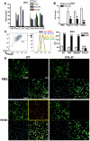
Figure 2
Figure 2. Dll1 expression expression is dependent upon type-I IFN.
(A) Gene expression of Dll1 on BMDMs from WT, TRIF−/−, MyD88−/−, or IFNαR−/− mice following stimulation with PolyI:C (10 µg/ml), LPS (1 μg/ml), CpG (1μM), H1N1 (MOI = 10), recombinant (r) IFN-α (20 Units), or rIFN-β (20 Units) for 6 hours. *P<0.05, **P<0.01 compared with WT mice. (B) The gene expression level of Dll1 on BMDMs pretreated with anti-mouse IFN-β Ab (2 µg/ml). *P<0.05, **P<0.01 compared with control (cont) Ab. (C) The level of Dll1 on BMDMs (CD11b+F4/80+) following stimulation with H1N1 (MOI = 10) for 24 hours determined by flow cytometry. **P<0.01 compared with PBS-treated BMDMs. (D) Confocal immunofluorescent examination of BMDMs stimulated with H1N1 (MOI = 10) between WT and IFNαR−/− mice. F4/80: green, Dll1: red, DAPI: blue. Original magnification, ×400. Data shown are mean ± SEM and are a representative experiment of 3 independent experiments. Each point represents at least 3 mice per group.
Figure 3
Figure 3. RIG-I- and JAK/STAT-dependent Dll1 induction in BMDMs.
(AC) BMDMs were transfected with RIG-I siRNA or control siRNA. The cells were then incubated with H1N1 for 24 hours (A, B) or for 6 hours (C), and the production of IFN-α (A) and IFN-β (B) was determined by ELISA, and the expression of Dll1 gene was measured by quantitative real-time PCR (C). **P<0.01 compared with control siRNA treated BMDMs. N.D.  =  not detectable (D) BMDMs were stimulated with PolyI:C (10 µg/ml), LPS (1 µg/ml), H1N1 (MOI = 10), or rIFN-β (20 Units) for 6 hours, and then expression of STAT1, phosphorylated (p)STAT1, STAT2, pSTAT2, and Dll1 were measured by western blotting. GAPDH was used as a loading control. (E, F) BMDMs were stimulated with PolyI:C (10 µg/ml), H1N1 (MOI = 10), or rIFN-β (20 Units) for 6 hours, and then Dll1 gene expression was analyzed by quantitative real-time PCR. (E) Dll1 expression on BMDMs between WT and STAT1−/− mice. **P<0.01 compared with WT (129S6) mice. (F) Dll1 expression on BMDMs between DMSO and JAK-I inhibitor treatment (10 µM). **P<0.01 compared with DMSO treatment. Data shown are mean ± SEM and are a representative experiment of 3 independent experiments. Each point represents at least 4 mice per group.
Figure 4
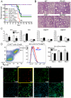
Figure 4. IFNαR
/ mice showed higher mortality with impaired Dll1 expression during influenza virus infection. (A) Survival rate in WT (black), IFNαR−/− (red), TRIF−/− (green), and MyD88−/− (blue) mice. Mice were inoculated intranasally with PBS (dotted line) or H1N1 at 1×10⁴ PFU (solid line) per each group of mice. Results are expressed as the percentage of survival from 10 individual mice per group. **P<0.01 compared with WT mice (B) Histological appearance of lungs from WT, IFNαR−/−, TRIF−/−, MyD88−/− mice at 8 days post-infection of influenza virus (C) Quantitative real-time PCR was performed to measure the transcript levels of Notch ligands at Day 4 and Day 8 after inoculation of influenza virus. *P<0.05 compared with WT mice (D) The level of Dll1 in lung macrophages (CD11b+F4/80+) from WT or IFNαR−/− mice was determined by flow cytometry at Day 4 and Day 8 after inoculation of influenza virus. *P<0.05, **P<0.01 compared with WT mice. (E) Confocal immunofluorescent examination of influenza infected lungs at 8 days post-infection, Dll1+ cells (red) merged with F4/80+ cells (green) in WT and IFNαR−/− mice. Blue staining indicates DAPI. Original magnification,×200. Shown are representative sections from 1 mouse of 4 per group. Data shown indicate mean ± SEM and are from a representative experiment of 3 independent experiments. Each time point represents at least 4 mice per group.
Figure 5
Figure 5. Macrophage is required for protection against influenza virus.
Liposome-DMDP or liposome alone (20 µl/dose) was intranasally administrated to mice on day 1 and day 4 after influenza virus challenge. (A) Survival rate of WT mice treated with either control liposome (black) or liposome-DMDP (red) after intranasal injection with PBS only (dotted line) or H1N1 (solid line). ***P<0.001 compared with H1N1 group treated with control liposome. (BD) Viral load in WT mice treated with either control liposome or liposome-DMDP at 2 and 7 days after infection of influenza virus (1×10⁴ PFU). (B) TCID50; Results are shown in log10 scale per lobe. M1 (C) and NS (D) H1N1 viral mRNA. Results are expressed as RNA copies normalized to GAPDH expression levels, as determined by real-time PCR. *P<0.05 compared with control liposome group. (E) Cellular cytospin appearance of bronchoalveolar lavage (BAL) harvested from mice treated with either control liposome or liposome-DMDP at day 2 and 7 after influenza virus infection stained with Giemsa stain. Red arrows show macrophages. Original magnification, ×1000. (F) The number of cells harvested from BAL. *P<0.05 compared with control liposome group. (G) Quantitative real-time PCR (Taqman) was performed to measure the transcript levels of Dll1 at day 2 and 7 after inoculation of influenza virus. *P<0.05 compared with control liposome group. (H) IFN-γ production from whole lungs of WT mice treated with either control liposome or liposome-DMDP at day 2 and 7 after influenza virus infection. Cytokine levels of IFN-γ were measured using a Luminex system. *P<0.05 compared with control liposome group. Data shown indicate mean ± SEM and are from a representative experiment of 2 independent experiments. Each time point represents 5 mice per group.
Figure 6
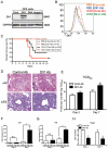
Figure 6. Passive immunization using Abs against Dll1 abrogates survival rate, lung pathology, and viral load.
(A) Western blot analysis showed the specificity of polyclonal rabbit anti-Dll1 using OP9 cells transfected with various Notch ligands. (B) The level of Dll1 in lung macrophages (CD11b+F4/80+) was determined with flow cytometry using an Ab against Dll1 at day7 post infection; PBS treated mice with either control Abs (Orange) or Abs directed against Dll1 (Blue), H1N1-infected mice with either control Abs (Red) or Abs directed against Dll1 (Green). (C) Survival rate of WT mice treated with either control Abs (black) or Abs directed against Dll1 (red) after intranasal injection with PBS only (dotted line) or H1N1 (solid line). **P<0.01 compared with control Ab group. (D) Histological appearance of lungs isolated from WT mice treated with either control Abs or Dll1 Abs at day 2 and 7 after influenza virus infection. H&E staining. Original magnification, ×40; ×200. (E–G) Viral load in WT mice treated with control Abs or anti-Dll1 Abs at 2 and 7 days after infection of influenza virus (1×10⁴ PFU), measured by TCID50 (E) and, M1 (F) and NS (G) H1N1 viral mRNA. Viral mRNAs are expressed as RNA copies normalized to GAPDH expression levels, as determined by real-time PCR. (H) mRNA expression of Hes1 in mice treated with control Abs or anti-Dll1 Abs 2 and 7 days after infection of influenza virus. *P<0.05 compared with control Ab group. Data shown indicate mean ± SEM and are from a representative experiment of 3 independent experiments. Each time point represents 5 mice per group.
Figure 7
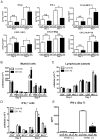
Figure 7. Blockade of Dll1 modulates immune response during influenza virus infection.
(A) Protein levels of cytokines and chemokines in whole lungs isolated from WT mice treated with either control Abs or anti-Dll1 Abs at day 2 and 7 after influenza virus infection using a Luminex or ELISA system. *P<0.05 compared with control Ab group. (B) FACS analysis of lung macrophage (MØ; CD11b+F4/80+), neutrophil (PMN; Gr-1high), myeloid DC (mDC; CD11b+CD11c+), and plasmacytoid DC (pDC; B220+CD11c+) isolated from influenza challenged mice at day 2 and 7. *P<0.05 compared with control Ab group. (C) FACS analysis of lung T cell (CD4+, CD8+) and NK cell (NK1.1+) isolated from influenza challenged mice at day 2 and 7. (D) FACS analysis of intracellular staining of CD4+ cells, CD8+ cells, NK1.1+ cells for IFN-γ. *P<0.05 compared with control Ab group. (E) Whole cells from the draining lymph nodes of WT mice treated with either control Abs or anti-Dll1 Abs at day 7 after influenza virus infection were restimulated in vitro with H1N1 for 48 hours. Cytokine level of IFN-γ was measured using a Luminex system. *P<0.05 compared with control Ab group. Data shown indicate mean ± SEM and are from a representative experiment of 3 independent experiments. Each time point indicates at least 4–5 mice per group.
Figure 8
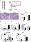
Figure 8. Blocking of Notch signaling abrogates survival rate, lung pathology, and viral load.
GSI (10 nM; 50 µl volume) was intranasally administrated to mice on day 1 and day 4 after influenza virus challenge. 20% DMSO in 50 µl was used as a control for GSI. (A) Survival rate of WT mice treated with either control DMSO (black) or GSI (red) after intranasal injection with PBS only (dotted line) or H1N1 (solid line). **P<0.01 compared with H1N1 injection group treated with DMSO (B) Histological appearance of lungs isolated from WT mice treated with either control DMSO or GSI at day 7 after influenza virus infection stained with H&E. Original magnification, ×100. (C–E) Viral load in WT mice treated with DMSO or GSI at 2 and 7 days after infection of influenza virus (1×10⁴ PFU) measured by TCID50 (C); Results are shown in log10 scale per lobe. H1N1 viral mRNAs, M1 (D) and NS (E), were measured. Results are expressed as RNA copies normalized to GAPDH expression levels, as determined by real-time PCR. **P<0.01 compared with H1N1 injection group treated with DMSO. (F) mRNA exprerssion of Hes1 from whole lungs. (G) FACS analysis of intracellular staining of CD4+ cells, CD8+ cells, NK1.1+ cells for IFN-γ. (H) Cytokine level of IFN-γ from whole lungs was measured using a Luminex system. *P<0.05 compared with H1N1 injection group treated with DMSO. Data shown indicate mean ± SEM and are from a representative experiment of 2 independent experiments. Each time point represents 45 mice per group.
Figure 9
Figure 9. Activation of IFN-γ from lung T cells by lung macrophages during immune responses.
(A, B) Lung CD4+ (A) or CD8+ (B) T cells were isolated from influenza virus challenged WT mice and stimulated with H1N1-pulsed lung-derived macrophages from either WT or IFNαR−/− mice. Cells were co-cultured with plate-coated rDll1 (2.5 µg/ml) or PBS control. (C, D) Lung CD4+ (C) or CD8+ (D) T cells were isolated from influenza virus challenged WT mice and stimulated with H1N1-pulsed lung-derived macrophages from either WT or IFNαR−/− mice. Cells were co-cultured with control IgG or anti-Dll1 Ab (20 µg/ml). Cytokine level of IFN-γ was measured using a Luminex system. Data shown are mean ± SEM and are from a representative experiment of 3 independent experiments. Each time point represents 4 mice per group. *P<0.05, ** P<0.01.

References

    1. Palese P. Influenza: old and new threats. Nat Med. 2004;10:S82–87. - PubMed
    1. Webby RJ, Webster RG. Are we ready for pandemic influenza? Science. 2003;302:1519–1522. - PubMed
    1. Swedish KA, Conenello G, Factor SH. First Season of 2009 H1N1 Influenza. Mt Sinai J Med. 2010;77:103–113. - PubMed
    1. Le Goffic R, Balloy V, Lagranderie M, Alexopoulou L, Escriou N, et al. Detrimental contribution of the Toll-like receptor (TLR)3 to influenza A virus-induced acute pneumonia. PLoS Pathog. 2006;2:e53. - PMC - PubMed
    1. Kumar H, Kawai T, Akira S. Pathogen recognition in the innate immune response. Biochem J. 2009;420:1–16. - PubMed

"V体育2025版" Publication types

MeSH terms