Skip to main page content
U.S. flag

An official website of the United States government

Dot gov

The . gov means it’s official VSports app下载. Federal government websites often end in . gov or . mil. Before sharing sensitive information, make sure you’re on a federal government site. .

Https

The site is secure. The https:// ensures that you are connecting to the official website and that any information you provide is encrypted and transmitted securely. V体育官网.

. 2012 Jan;33(1):156-63.
doi: 10.1093/carcin/bgr237. Epub 2011 Nov 9.

Induction of NAD(P)H-quinone oxidoreductase 1 by antioxidants in female ACI rats is associated with decrease in oxidative DNA damage and inhibition of estrogen-induced breast cancer

Affiliations

Induction of NAD(P)H-quinone oxidoreductase 1 by antioxidants in female ACI rats is associated with decrease in oxidative DNA damage and inhibition of estrogen-induced breast cancer (V体育2025版)

Bhupendra Singh et al. Carcinogenesis. 2012 Jan.

Abstract (V体育平台登录)

Exact mechanisms underlying the initiation and progression of estrogen-related cancers are not clear. Literature, evidence and our studies strongly support the role of estrogen metabolism-mediated oxidative stress in estrogen-induced breast carcinogenesis. We have recently demonstrated that antioxidants vitamin C and butylated hydroxyanisole (BHA) or estrogen metabolism inhibitor α-naphthoflavone (ANF) inhibit 17β-estradiol (E2)-induced mammary tumorigenesis in female ACI rats. The objective of the current study was to identify the mechanism of antioxidant-mediated protection against E2-induced DNA damage and mammary tumorigenesis. Female ACI rats were treated with E2 in the presence or absence of vitamin C or BHA or ANF for up to 240 days. Nuclear factor erythroid 2-related factor 2 (NRF2) and NAD(P)H-quinone oxidoreductase 1 (NQO1) were suppressed in E2-exposed mammary tissue and in mammary tumors after treatment of rats with E2 for 240 days. This suppression was overcome by co-treatment of rats with E2 and vitamin C or BHA. Time course studies indicate that NQO1 levels tend to increase after 4 months of E2 treatment but decrease on chronic exposure to E2 for 8 months. Vitamin C and BHA significantly increased NQO1 levels after 120 days VSports手机版. 8-Hydroxydeoxyguanosine (8-OHdG) levels were higher in E2-exposed mammary tissue and in mammary tumors compared with age-matched controls. Vitamin C or BHA treatment significantly decreased E2-mediated increase in 8-OHdG levels in the mammary tissue. In vitro studies using silencer RNA confirmed the role of NQO1 in prevention of oxidative DNA damage. Our studies further demonstrate that NQO1 upregulation by antioxidants is mediated through NRF2. .


V体育ios版 - Figures
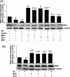
Fig. 1.
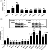
Fig. 1.
Antioxidants reverse E2-mediated decrease in NQO1 expression. Female ACI rats were treated with E2, BHA, BHA + E2, Vit C, Vit C + E2, ANF and ANF + E2 for 240 days as described in the Materials and methods section. At the end of the experiment, mammary tumors and mammary tissues were collected and used for western blot analysis for NQO1. Intensities of the bands were quantified and normalized to α-tubulin. Fold change compared to respective age-matched control mammary tissue was calculated from at least three different animals in each group. (A) NQO1 protein expression is significantly decreased in E2-treated mammary tumor and mammary tissues. Antioxidants BHA and Vit C increased NQO1 protein expression and its level remains unchanged after treatment with ANF compared to age-matched controls. The bar graph represents NQO1 protein fold change (mean ± SEM) in mammary tumors and mammary tissues from at least three different animals compared to age-matched control mammary tissues. (B) E2 (10 nM) treatment of MCF-10A cells for 24 h decreased NQO1 protein expression, whereas antioxidants BHA (250 μM) and Vit C (1 mM) significantly increased NQO1 protein levels. The bar graph represents NQO1 protein fold change (mean ± SEM) in MCF-10A from at least three different replicates compared to vehicle-treated MCF-10A. Significance was determined by analysis of variance and least significant difference post-hoc analysis. The symbols a, b and c indicate a P value <0.05 between specific treatment group and control, between specific treatment group and E2-treated mammary and between specific treatment group and mammary tumor, respectively.
Fig. 2.
Fig. 2.
E2 inhibits, whereas BHA and Vit C increase NQO1 in time-dependent fashion. Female ACI rats were treated with E2, BHA, BHA + E2, Vit C and Vit C + E2 for 7 to 240 days as described in the Materials and methods section. At the end of the experiment, mammary tumor and mammary tissues were collected. Tissues were homogenized and approximately 80 μg protein was used for western blot analysis. Intensities of the bands were quantified and normalized to α-tubulin. (A) NQO1 protein expression in mammary tumors and E2-treated mammary tissues after 7, 15, 120 and 240 days of treatment. The bar graph represents NQO1 protein fold change (mean ± SEM) in mammary tumors and mammary tissues from at least three different animals compared to age-matched control mammary tissues. (B) NQO1 protein expression in BHA and BHA + E2-treated group mammary tissues after 7, 15, 120 and 240 days of treatment. The bar graph represents NQO1 protein fold change (mean ± SEM) in mammary tissues from at least three different animals compared to age-matched control mammary tissues. (C) NQO1 protein expression in Vit C and Vit C + E2-treated group mammary tissues after 7, 15, 120 and 240 days of treatment. The bar graph represents NQO1 protein fold change (mean ± SEM) in mammary tissues from at least three different animals compared to age-matched control mammary tissues. ‘*’ indicates P value <0.05 compared to respective control mammary tissue.
Fig. 3.
Fig. 3.
Antioxidant-mediated change in NQO1 protein expression is tissue specific. Female ACI rats were treated with E2, BHA, BHA + E2, Vit C and Vit C + E2 for 240 days as described in the Materials and methods section. At the end of the experiment, kidney, heart, lung, liver, brain, spleen, thymus and uterus tissues were collected. Western blot analysis was carried out to investigate NQO1 protein expression in different tissues. NQO1 protein fold changes in respective organ from at least three different animals compared to respective control organ are represented at the top of each blot. ‘*’ indicates P value <0.05 compared to control mammary tissue.
Fig. 4.
Fig. 4.
Antioxidants upregulate NQO1 via NRF2 -dependent pathway. (A) Female ACI rats were treated with E2, Vit C, Vit C + E2, BHA and BHA + E2 for 240 days as described in the Materials and methods section. At the end of the experiment, mammary tumor and mammary tissues were collected. Western blot analysis was carried out to investigate NRF2 and NQO1 protein expression in mammary tumor and mammary tissues. The bar graph represents NRF2 and NQO1 protein fold change (mean ± SEM) in mammary tumors and mammary tissues from at least three different animals compared to age-matched control mammary tissues. ‘*’ indicates P value <0.05 for NRF2 protein expression compared to respective control mammary. ‘**’ indicates a P value <0.05 for NQO1 protein expression compared to respective control mammary. (B) MCF-10A cells were transfected with 20 nmol/l of scrambled small interfering RNA, siNQO1 or siNRF2 for 48 h and western blot analysis was carried out using NRF2 antibody. Same membrane was reprobed with NQO1 and α-tubulin antibody. (C) MCF-10A cells were treated with Vit C (1 mM) or BHA (250 μM) in the presence or absence of E2 (10 nM) or vehicle for 2 h, fixed with formaldehyde, cross-linked and the chromatin sheared. The chromatin was immunoprecipitated with NRF2 antibody. NRF2 binding to NQO1 promoter was analyzed by real-time PCR with specific primers for the human NQO1 ARE region. The ARE region of the NQO1 promoter was also amplified from 3 μl of purified soluble chromatin before immunoprecipitation to show input DNA. Representative ChIP agarose gels and real-time PCR Ct values from three independent experiments are shown (ND, not detected).
Fig. 5.
Fig. 5.
Antioxidants inhibit E2-mediated oxidative DNA damage. Female ACI rats were treated with E2, Vit C, Vit C + E2, BHA, BHA + E2, ANF and ANF + E2 for 240 days as described in the Materials and methods section. 8-OHdG levels were measured in mammary tumors and mammary tissues from animals in each of these groups as well as in MCF-10A cells treated with above chemicals and dicumarol. siNQO1- or siNRF2 -transfected MCF-10A cells were also treated with E2 (50 nM) for 48 h and 8-OHdG levels quantified. (A) Levels of 8-OHdG in mammary tumors and mammary tissues from different treatment groups. These data are reported as an average of values obtained for at least six different animals ± SEM. (B) Levels of 8-OHdG in MCF-10A cells treated with Vit C, BHA, ANF and dicumarol in presence or absence of E2 and in NQO1 and NRF2 knockdown MCF-10A cells. Inhibition of NQO1 and NRF2 protein expression in siNQO1 and siNRF2-transfected MCF-10A cells is shown in inset. Significance was determined by analysis of variance and least significant difference post-hoc analysis. The symbols a, b, c, d, e, f and g indicate a P value <0.05 between specific treatment group and control, between specific treatment group and E2 treatment, between specific treatment group and mammary tumor, between siNQO1 or siNRF2 and scrambled, between siNQO1 and siNQO1 + E2, between siNRF2 and siNRF2 + E2 and between dicumarol and dicumarol + E2, respectively.

References

    1. Bhat HK, et al. Critical role of oxidative stress in estrogen-induced carcinogenesis. Proc. Natl. Acad. Sci. USA. 2003;100:3913–3918. - PMC - PubMed
    1. Cavalieri EL, et al. Molecular origin of cancer: catechol estrogen-3,4-quinones as endogenous tumor initiators. Proc. Natl. Acad. Sci. USA. 1997;94:10937–10942. - "VSports在线直播" PMC - PubMed
    1. Henderson BE, et al. Hormonal carcinogenesis. Carcinogenesis. 2000;21:427–433. - PubMed
    1. Mense SM, et al. Estrogen-induced breast cancer: alterations in breast morphology and oxidative stress as a function of estrogen exposure. Toxicol. Appl. Pharmacol. 2008;232:78–85. - PMC - PubMed
    1. Mense SM, et al. Vitamin C and alpha-naphthoflavone prevent estrogen-induced mammary tumors and decrease oxidative stress in female ACI rats. Carcinogenesis. 2009;30:1202–1208. - PMC - PubMed

Publication types (VSports手机版)

MeSH terms