Skip to main page content (VSports app下载)
U.S. flag

An official website of the United States government

Dot gov

The . gov means it’s official. Federal government websites often end in VSports app下载. gov or . mil. Before sharing sensitive information, make sure you’re on a federal government site. .

Https

The site is secure. The https:// ensures that you are connecting to the official website and that any information you provide is encrypted and transmitted securely V体育官网. .

. 2012 Jan;22(1):168-77.
doi: 10.1038/cr.2011.177. Epub 2011 Nov 8.

The metabolome of induced pluripotent stem cells reveals metabolic changes occurring in somatic cell reprogramming

Affiliations

"VSports注册入口" The metabolome of induced pluripotent stem cells reveals metabolic changes occurring in somatic cell reprogramming

Athanasia D Panopoulos et al. Cell Res. 2012 Jan.

Abstract

Metabolism is vital to every aspect of cell function, yet the metabolome of induced pluripotent stem cells (iPSCs) remains largely unexplored. Here we report, using an untargeted metabolomics approach, that human iPSCs share a pluripotent metabolomic signature with embryonic stem cells (ESCs) that is distinct from their parental cells, and that is characterized by changes in metabolites involved in cellular respiration. Examination of cellular bioenergetics corroborated with our metabolomic analysis, and demonstrated that somatic cells convert from an oxidative state to a glycolytic state in pluripotency. Interestingly, the bioenergetics of various somatic cells correlated with their reprogramming efficiencies VSports手机版. We further identified metabolites that differ between iPSCs and ESCs, which revealed novel metabolic pathways that play a critical role in regulating somatic cell reprogramming. Our findings are the first to globally analyze the metabolome of iPSCs, and provide mechanistic insight into a new layer of regulation involved in inducing pluripotency, and in evaluating iPSC and ESC equivalence. .


Figures (VSports最新版本)

Figure 1
Figure 1
The metabolome profiles of iPSCs versus ESCs. Heat maps of metabolite features (> 5 000) in the indicated iPSC and ESC lines grown in chemically defined conditions at early (left panel) and late passage (right panel). The percentage of metabolite feature differences in iPSCs compared to ESCs is indicated below each respective heat map. Biological duplicates (e.g., (a) vs (b)) and experimental duplicates (e.g., (a) vs (a)) were performed.
Figure 2
Figure 2
Metabolic differences between iPSCs and ESCs reveal additional pathways important for reprogramming. (A) Metabolites that differ between ESCs and iPSCs (grown in chemically defined conditions and isolated at late passage) as identified by accurate mass and MS/MS data. Fold values comparing the median integrated peak intensities are shown. (B) Integrated peak intensities for a sample of metabolites identified in ESCs, FiPSCs and parental fibroblast somatic populations. (C) dFib-OCT4GFP cells infected with retroviruses encoding KLF-4, OCT4, SOX2 and c-MYC (KOSM) were grown in ESC medium in the presence or absence of arachidonic acid (AA, 0.1 μM), S-adenosyl methionine (SAM, 0.5 mM), or ethanol (EtOH) alone as a control. The number of GFP-positive colonies were counted ∼16 days after the initial infection, and are plotted for each condition relative to controls. Error bars depict the SEM. *P< 0.05.
Figure 3
Figure 3
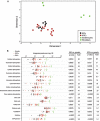
Pluripotent cells have a distinct metabolic signature characterized by changes in metabolites involved in cellular respiration. (A) Two-dimensional representation of the XCMS matrix of retention time, m/z, and feature intensity values using a multidimensional scaling (MDS) plot for ESCs, iPSCs (derived from human keratinocytes and fibroblasts) and their somatic cells of origin. Data points for cell populations producing similar features are closer to one another than data points for cell populations producing more dissimilar features. Each cell line was analyzed by at least two biological and experimental replicates. (B) Quantification of metabolites from ESCs, iPSCs and somatic cell populations of origin (keratinocytes and fibroblasts). Data points and bars represent the integrated peak intensity for each sample and the median intensity values. Fold values indicate the difference in integrated peak intensity (median value) for selected metabolites in ESCs and iPSCs relative to somatic populations. Identification is based on accurate mass and MS/MS data. Each cell line was analyzed by at least two biological and experimental replicates.
Figure 4
Figure 4
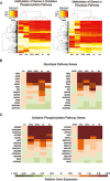
Genes involved in glycolysis and oxidative phosphorylation undergo changes in methylation and gene expression following the transition from a somatic to a pluripotent state. (A) Levels of epigenetic changes occurring in genes regulating glycolysis and oxidative phosphorylation (see Materials and Methods). Heatmap and hierarchical clustering results of keratinocytes, fibroblasts, their respective iPSC lines (KiPS and FiPS) and ESC lines (H1and H9) using methylation patterns at CpG sites containing a change in methylation in at least one iPSC line. Clustering was performed on a dissimilarity matrix with values equal to the complement of the Pearson's correlation for each pair. (B-C) A selected number of genes involved in glycolysis (B) or oxidative phosphorylation (C), that demonstrated differentially methylated sites between somatic cells and their respective iPSC counterparts, were measured for expression changes by real-time PCR (see Supplementary information, Tables S1 and S2). A heatmap of gene expression in iPSC lines and ESC controls (H1 and H9) relative to their somatic cell of origin is shown.
Figure 5
Figure 5
Reprogramming induces a bioenergetic conversion from an oxidative to a glycolytic state. (A) Basal oxygen consumption rate (OCR, indicative of mitochondrial oxidative phosphorylation) plotted versus basal extracellular acidification rate (ECAR, representing glycolysis) for IMR90 fibroblasts, keratinocytes and their respective iPSCs (FiPS4F5, KiPS4FB). Results represent the average of four independent experiments performed in triplicate. (B) OCR/ECAR ratios of H1 ESCs, HUVECs, IMR90 and BJ fibroblasts, and iPSCs from HUVECs (Huv-iPS), keratinocytes (KiPS) or IMR90 fibroblasts (FiPS) are shown. Results were determined from the average of five independent experiments performed in triplicate. (C) dFib-OCT4GFP cells were infected with retroviruses encoding KLF-4, OCT4, SOX2 and c-MYC (KOSM). Equivalent numbers of KOSM-infected cells were plated and grown in ESC medium in the presence or absence of 2-deoxy-D-glucose (2-DG, 1 mM) or D-fructose-6-phosphate (F6P, 5 μM). GFP-positive colonies were numerated at ∼day 16 after the initial infection. The number of GFP-positive colonies for each condition relative to controls are shown. (D) dFib-OCT4GFP cells were treated with 2-deoxy-D-glucose (2-DG, 1 mM) or D-fructose-6-phosphate (F6P, 5 μM) for 48 h, and ECAR and OCR values relative to media only controls determined. All OCR and ECAR values were normalized to cell number. Error bars depict the SEM. *P < 0.05.

References

    1. Takahashi K, Yamanaka S. Induction of pluripotent stem cells from mouse embryonic and adult fibroblast cultures by defined factors. Cell. 2006;126:663–676. - "V体育安卓版" PubMed
    1. Takahashi K, Tanabe K, Ohnuki M, et al. Induction of pluripotent stem cells from adult human fibroblasts by defined factors. Cell. 2007;131:861–872. - PubMed
    1. Yu J, Vodyanik MA, Smuga-Otto K, et al. Induced pluripotent stem cell lines derived from human somatic cells. Science. 2007;318:1917–1920. - PubMed
    1. Stadtfeld M, Hochedlinger K. Induced pluripotency: history, mechanisms, and applications. Genes Dev. 2010;24:2239–2263. - PMC - PubMed
    1. Panopoulos AD, Ruiz S, Izpisua Belmonte JC. iPSCs: induced back to controversy. Cell Stem Cell. 2011;8:347–348. - PubMed

V体育官网 - Publication types

MeSH terms