<u dropzone="RfZPb5qj"></u> Skip to main page content
U.S. flag

An official website of the United States government

Dot gov

The VSports app下载. gov means it’s official. Federal government websites often end in . gov or . mil. Before sharing sensitive information, make sure you’re on a federal government site. .

Https

The site is secure V体育官网. The https:// ensures that you are connecting to the official website and that any information you provide is encrypted and transmitted securely. .

. 2012 Feb;40(3):981-95.
doi: 10.1093/nar/gkr818. Epub 2011 Oct 3.

"VSports最新版本" The acetylation of transcription factor HBP1 by p300/CBP enhances p16INK4A expression

Affiliations

"V体育ios版" The acetylation of transcription factor HBP1 by p300/CBP enhances p16INK4A expression

Weibin Wang et al. Nucleic Acids Res. 2012 Feb.

Abstract

HBP1 is a sequence-specific DNA-binding transcription factor with many important biological roles. It activates or represses the expression of some specific genes during cell growth and differentiation. Previous studies have exhibited that HBP1 binds to p16(INK4A) promoter and activates p16(INK4A) expression. We found that trichostatin A (TSA), an inhibitor of HDAC (histone deacetylase), induces p16(INK4A) expression in an HBP1-dependent manner. This result was drawn from a transactivation experiment by measuring relative luciferase activities of p16(INK4A) promoter with HBP1-binding site in comparison with that of the wild-type p16(INK4A) promoter by transient cotransfection with HBP1 into HEK293T cells and 2BS cells. HBP1 acetylation after TSA treatment was confirmed by immunoprecipitation assay. Our data showed that HBP1 interacted with histone acetyltransferase p300 and CREB-binding protein (CBP) and also recruited p300/CBP to p16(INK4A) promoter. HBP1 was acetylated by p300/CBP in two regions: repression domain (K297/305/307) and P domain (K171/419). Acetylation of Repression domain was not required for HBP1 transactivation on p16(INK4A). However, luciferase assay and western blotting results indicate that acetylation of P domain, especially K419 acetylation is essential for HBP1 transactivation on p16(INK4A) VSports手机版. As assayed by SA-beta-gal staining, the acetylation of HBP1 at K419 enhanced HBP1-induced premature senescence in 2BS cells. In addition, HDAC4 repressed HBP1-induced premature senescence through permanently deacetylating HBP1. We conclude that our data suggest that HBP1 acetylation at K419 plays an important role in HBP1-induced p16(INK4A) expression. .


"VSports手机版" Figures
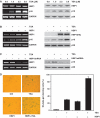
Figure 1.
Figure 1.
HBP1-induced p16INK4A expression was enhanced by TSA treatment. (A) 2BS cells were treated with TSA at different concentration (0, 1, 2.5, 5 μM) for 18 h. The mRNA levels of p16INK4A and HBP1 were determined by RT–PCR (left panel). The protein levels of HBP1 and p16INK4A were determined by western blot (right panel). (B) 2BS cells were stably infected with pITA-HBP1. After selection for 3 days, cells were treated with TSA at 1 μM for 18 h. mRNA (left panel) and protein (right panel) were extracted for analysis. (C) 2BS cells were transfected with pHBP1shRNA or pSuper.retro (as control). After selection for 3 days, cells were treated with TSA at 1 μM for 18 h. The left panel shows HBP1 knockdown efficiency determined by RT–PCR and the right panel shows the protein level of p16INK4A measured by western blot. (D) Young 2BS cells (PD20) were transfected with pITA-HBP1. After selected with puromycin for 3 days, cells were treated with TSA. Then the cells were stained for SA-β-gal (left panel). The percentage of cells positive for SA-β-gal in 2BS cells is shown in right panel. At least 300 cells were counted for each sample.
Figure 2.
Figure 2.
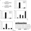
TSA increased HBP1 transactivation on p16INK4A by enhancing the DNA binding of HBP1 to the promoter region of p16INK4A. (A) The integrity of binding site is indispensible for HBP1 enhancing p16INK4A promoter. Schematic diagrams of the native p16INK4A promoter (NP) and its mutant promoter (MP) (left panel). The mutant promoter was constructed by changing one point (T to A) in HBP1-binding site in the background of native p16INK4A promoter. Luciferase assay was performed in cells transfected with NP, or MP and HBP1 to test p16INK4A mutant promoter (right panel). (B) HBP1-binding site is required for activating TSA-induced p16INK4A expression. Thirty hours after transfection with either pGL3-NP or pGL3-MP, 2BS cells were treated with TSA at 1 μM for 18 h. Relative luciferase activity was measured after TSA treatment. (C) The transactivation of HBP1 on p16INK4A is enhanced by TSA. Thirty hours after transfection with HBP1, cells were treated with TSA at 1 μM for 18 h. Then the cells were harvested for luciferase assay. (D) TSA loses its transactivation ability at the p16INK4A promoter when HBP1 was knocked-down with shRNA. Thirty hours after transfection with HBP1shRNA, cells were treated with TSA at 1 μM for 18 h. Then the cells were harvested for luciferase assay. (E) TSA enhances the DNA binding of HBP1 to the promoter region of p16INK4A in vivo. ChIP assay with antibody against HBP1 was performed in 2BS cells with or without TSA treatment at 1 μM for 18 h. Lanes with input chromatin were positive controls (lanes 2, 6). Lanes without chromatin or GAPDH antibody were used for negative controls (lanes 1, 3, 5, 7). At the bottom, anti-HBP1 western immunoblotting (IB) for HBP1 protein expression is shown.
Figure 3.
Figure 3.
HBP1 was acetylated within the cells. (A) Exogenous HBP1 was acetylated within the cells. 2BS cells transfected with HBP1(HA-tagged) were treated with TSA at 1 μM for 18 h. IP assay was carried out by using HA antibody and followed by western blot with acetyl-lysine antibody (left). IP assay was carried out by using acetyl-lysine antibody and followed by western blot with HA antibody (right). (B) Endogenous HBP1 was acetylated within the cells. 2BS cells were treated with TSA at 1 μM for 18 h. IP assay was carried out by using HBP1 antibody and followed by western blot with acetyl-lysine antibody.
Figure 4.
Figure 4.
HBP1 interacted with p300/CBP and recruits p300/CBP onto the promoter of p16INK4A. (A) Exogenous HBP1 interacts with p300/CBP. HEK293T cells transfected with HA-HBP1 were subjected to immunoprecipitation with anti-HA antibody. Immunoprecipitated complexes were used for western blot analysis with anti- p300, anti-CBP or anti-PCAF antibody, respectively (left). The interaction of HBP1 and p300/CBP was further confirmed by reciprocal analysis. IP was performed with anti-p300, anti-CBP or anti-PCAF antibody respectively and followed with western blot with anti-HA antibody (right). The bottom lanes were for determining IP efficiency. (B) Endogenous HBP1 interacted with p300/CBP. HEK293T cells were subjected to immunoprecipitation with anti-HBP1 antibody. Immunoprecipitated complexes were used for western blot analysis with anti-p300, anti-CBP or anti-PCAF antibody respectively. The bottom lanes were for determining IP efficiency. (C) Endogenous HBP1 cannot be acetylated when p300/CBP knockdown. HEK293T cells were transfected with p300siRNA and CBPsiRNA. The protein levels of p300 and CBP were determined by western blot (left panel). Then the cells were subjected to immunoprecipitation with anti-HBP1 antibody with or without TSA treatment. Immunoprecipitated complexes were used for western blot analysis with acetyl-lysine antibody (Right panel). (D) HBP1 is required for p300/CBP recruitment onto p16INK4A promoter when TSA was applied. HEK293T cells were transfected with HBP1shRNA. The protein level of HBP1 was determined by western blot (left panel). ChIP assay with anti-p300, anti-CBP and anti-PCAF antibody was performed in HEK293T cells after treated with TSA at 2.5 μM for 18 h with or without HBP1shRNA transfection. (E) TSA loses its transactivation ability at the p16INK4A promoter when p300/CBP knockdown. Thirty hours after transfection with p300/CBP siRNA, cells were treated with TSA at 2.5 μM for 18 h. Then the cells were harvested for luciferase assay.
Figure 5.
Figure 5.
HDAC4 represses HBP1 ability to induce senescence. (A) 2BS cells were stably infected with pITA-HBP1. After selection for 3 days, cells were transfected with pITA-HDAC4. The protein levels of HBP1 (HA), HDAC4 (Flag) and p16INK4A were measured by western blot (left panel). Then the cells were stained for SA-β-gal. The percentage of cells positive for SA-β-gal in 2BS cells is shown in right panel. At least 300 cells were counted for each sample. (B) 2BS cells were stably infected with pITA-HBP1. After selection for 3 days, cells were transfected with pSR-HDAC4shRNA. The protein levels of HBP1 (HA), HDAC4 and p16INK4A were measured by western blot (left panel). Then the cells were stained for SA-β-gal. The percentage of cells positive for SA-β-gal in 2BS cells is shown in right panel. At least 300 cells were counted for each sample.
Figure 6.
Figure 6.
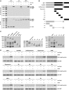
Wild-type HBP1 and mutants were acetylated by HAT of p300/CBP in vitro. (A) HBP1 was acetylated by HAT in vitro. At the top, purified GST-tagged HBP1 protein was incubated with HAT in the presence of Ac-CoA. The reaction products were separated by SDS–PAGE and immunoblotted with the acetyl-lysine antibody. At the bottom, the same nitrocellulose membrane as used in the in vitro acetylation assay described above was stripped and immunoblotted with anti-GST antibody. GST and GST-tagged p53 proteins were used for negative and positive controls, respectively. (B) Schematic diagram of wild-type HBP1 and associated mutants. (C) Expression of wild-type HBP1 and associated mutants proteins in E. coli. GST-tagged proteins were immunoprecipited with GST, then ran on SDS–PAGE and stained with silver. (D) HBP1 and some mutants (Repression domain, P domain) were acetylated by HAT in vitro. GST-tagged proteins from experiments shown in (B) were incubated with Ac-CoA and HAT in 30°C for 4 h. The reaction products were separated by SDS–PAGE and immunoblotted with the acetyl-lysine antibody. GST was as loading control.
Figure 7.
Figure 7.
The K297, K305, K307 of Repression domain and K171, K419 of P domain were acetylated by HAT of p300/CBP in vitro. (A) Schematic diagram of associated mutants. (B) Expression of associated mutants proteins in E. coli. GST-tagged proteins were immunoprecipited with GST, then ran on SDS–PAGE and stained with silver. (C) The K297, K305, K307 of Repression domain and K171, K419 of P domain are acetylated by HAT of p300/CBP in vitro. GST-tagged proteins from experiments shown in panel A were incubated with Ac-CoA and HAT in 30°C for 4 h. The reaction products were separated by SDS–PAGE and immunoblotted with the acetyl-lysine antibody. GST was as loading control.
Figure 8.
Figure 8.
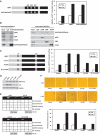
The acetylation of HBP1 at K419 was indispensible for HBP1-induced premature senescence. (A) HBP1 acetylation at K297/305/307 was not required for transactivation on p16INK4A. HEK293T cells were cotransfected with HBP1 or point mutants K297/305/307R and pGL3-p16INK4A promoter for 48 h. After transfection, cells were treated with TSA at 2.5 μM for 18 h. Then the cells were harvested for luciferase assay. (B) K297/305/307 was not required for interaction of HBP1 and p300/CBP. The whole cell extracts were subjected to immunoprecipitation with anti-HA antibody. Immunoprecipitated complexes were used for western blot analysis with anti-p300, anti-CBP or anti-PCAF antibody respectively. The bottom lanes were for determining IP efficiency. The interaction of HBP1 and p300/CBP was further confirmed by reciprocal analysis. (C) HBP1 acetylation at K419 was required for transactivation on p16INK4A. HEK293T cells were transfected with pGL3-p16INK4A promoter and HBP1 or mutants for 48 h. After transfection, cells were treated with TSA at 2.5 μM for 18 h. Then the cells were harvested for luciferase assay. (D) HBP1 acetylation at K419 was required for HBP1-induced p16INK4A expression. p16INK4A protein level in 2BS cells transfected with HBP1 and mutants for 5-7 days was determined by western blot. (E) HBP1 acetylation at K419 was required for the binding of HBP1 onto p16INK4A promoter. ChIP assay with antibody against HA was performed in HEK293T cells transfected with HBP1 or mutants. (F) HBP1 acetylation at K419 is required for HBP1-induced premature senescence. 2BS cells transfected with HBP1 or mutants with or without TSA treatment were stained for SA-β-gal (up panel). The percentage of cells positive for SA-β-gal is shown in low panel. At least 300 cells were counted for each sample. (G) p300/CBP knockdown attenuates the HBP1-induced p16INK4A gene activity. 2BS cells were stably infected with pITA-HBP1. After selection for 3 days, cells were transfected with p300/CBP siRNA and pGL3-p16INK4A promoter. After transfection, cells were treated with TSA at 1 μM for 18 h. Then the cells were harvested for luciferase assay. (H) p300/CBP knockdown attenuates the HBP1-induced senescence. 2BS cells were stably infected with pITA-HBP1. After selection for 3 days, cells were transfected with p300/CBP siRNA. After transfection, cells were treated with TSA at 1 μM for 18 h. Then the cells were stained for SA-β-gal.
Figure 8.
Figure 8.
The acetylation of HBP1 at K419 was indispensible for HBP1-induced premature senescence. (A) HBP1 acetylation at K297/305/307 was not required for transactivation on p16INK4A. HEK293T cells were cotransfected with HBP1 or point mutants K297/305/307R and pGL3-p16INK4A promoter for 48 h. After transfection, cells were treated with TSA at 2.5 μM for 18 h. Then the cells were harvested for luciferase assay. (B) K297/305/307 was not required for interaction of HBP1 and p300/CBP. The whole cell extracts were subjected to immunoprecipitation with anti-HA antibody. Immunoprecipitated complexes were used for western blot analysis with anti-p300, anti-CBP or anti-PCAF antibody respectively. The bottom lanes were for determining IP efficiency. The interaction of HBP1 and p300/CBP was further confirmed by reciprocal analysis. (C) HBP1 acetylation at K419 was required for transactivation on p16INK4A. HEK293T cells were transfected with pGL3-p16INK4A promoter and HBP1 or mutants for 48 h. After transfection, cells were treated with TSA at 2.5 μM for 18 h. Then the cells were harvested for luciferase assay. (D) HBP1 acetylation at K419 was required for HBP1-induced p16INK4A expression. p16INK4A protein level in 2BS cells transfected with HBP1 and mutants for 5-7 days was determined by western blot. (E) HBP1 acetylation at K419 was required for the binding of HBP1 onto p16INK4A promoter. ChIP assay with antibody against HA was performed in HEK293T cells transfected with HBP1 or mutants. (F) HBP1 acetylation at K419 is required for HBP1-induced premature senescence. 2BS cells transfected with HBP1 or mutants with or without TSA treatment were stained for SA-β-gal (up panel). The percentage of cells positive for SA-β-gal is shown in low panel. At least 300 cells were counted for each sample. (G) p300/CBP knockdown attenuates the HBP1-induced p16INK4A gene activity. 2BS cells were stably infected with pITA-HBP1. After selection for 3 days, cells were transfected with p300/CBP siRNA and pGL3-p16INK4A promoter. After transfection, cells were treated with TSA at 1 μM for 18 h. Then the cells were harvested for luciferase assay. (H) p300/CBP knockdown attenuates the HBP1-induced senescence. 2BS cells were stably infected with pITA-HBP1. After selection for 3 days, cells were transfected with p300/CBP siRNA. After transfection, cells were treated with TSA at 1 μM for 18 h. Then the cells were stained for SA-β-gal.

References

    1. Berasi SP, Xiu M, Yee AS, Paulson KE. HBP1 repression of the p47phox gene: cell cycle regulation via the NADPH oxidase. Mol. Cell. Biol. 2004;24:3011–3024. - PMC - PubMed
    1. Paulson KE, Rieger-Christ K, McDevitt MA, Kuperwasser C, Kim J, Unanue VE, Zhang X, Hu M, Ruthazer R, Berasi SP, et al. Alterations of the HBP1 transcriptional repressor are associated with invasive breast cancer. Cancer Res. 2007;67:6136–6145. - PubMed
    1. Lavender P, Vandel L, Bannister AJ, Kouzarides T. The HMG-box transcription factor HBP1 is targeted by the pocket proteins and E1A. Oncogene. 1997;14:2721–2728. - PubMed
    1. Lemercier C, Duncliffe K, Boibessot I, Zhang H, Verdel A, Angelov D, Khochbin S. Involvement of retinoblastoma protein and HBP1 in histone H1(0) gene expression. Mol. Cell. Biol. 2000;20:6627–6637. - PMC - PubMed
    1. Shih HH, Tevosian SG, Yee AS. Regulation of differentiation by HBP1, a target of the retinoblastoma protein. Mol. Cell. Biol. 1998;18:4732–4743. - PMC - PubMed

Publication types

MeSH terms