Skip to main page content
U.S. flag

An official website of the United States government

Dot gov

The VSports app下载. gov means it’s official. Federal government websites often end in . gov or . mil. Before sharing sensitive information, make sure you’re on a federal government site. .

Https

The site is secure. The https:// ensures that you are connecting to the official website and that any information you provide is encrypted and transmitted securely. V体育官网.

. 2011 Oct;2(5):298-309.
doi: 10.1007/s12672-011-0084-4.

PI3K, Erk signaling in BMP7-induced epithelial-mesenchymal transition (EMT) of PC-3 prostate cancer cells in 2- and 3-dimensional cultures

Affiliations

PI3K, Erk signaling in BMP7-induced epithelial-mesenchymal transition (EMT) of PC-3 prostate cancer cells in 2- and 3-dimensional cultures

Minyoung Lim et al. Horm Cancer. 2011 Oct.

"V体育2025版" Abstract

We reported previously that bone morphogenetic protein 7 (BMP7) could induce epithelial-mesenchymal transition (EMT) in PC-3 prostate cancer cells grown in tissue culture plates. In this study, we examined BMP7-induced morphological and molecular expression changes that are characteristic of EMT using these cells under both two- (2D) and three-dimensional (3D) culture conditions VSports手机版. Filamentous outgrowths from spheroid structures that were formed from PC-3 cells in 3D cultures were strikingly evident when the spheroids were exposed to extracellular BMP7. This morphological change in 3D was accompanied by down-regulation of E-cadherin, which is an essential adhesion molecule for the integrity of epithelial phenotype. Invasiveness of the cancer cells was significantly enhanced with BMP7 treatment along with activation and up-regulation of proteases such as MMP1, MMP13, and urokinase plasminogen activator. Signal transduction of EMT conversion was examined by the use of certain pathway-specific inhibitors. Of the chemical inhibitors tested, inhibitors of PI3 kinase and Erk were found to suppress BMP-induced morphological changes both in 2D and 3D conditions. These results suggest that, besides the Smad signaling pathways, BMP-induced activation of PI3K and Erk contribute to EMT morphologic conversion of the PC-3 prostate cancer cells. Together, the results support the notion that the complexity of EMT may be better evaluated in terms of both spatial and temporal processes in 3D cell culture models that are physiologically more relevant than the cell growth in tissue culture plates. .


Conflict of interest statement

The authors declare that they have no conflict of interest.

Figures (V体育官网)
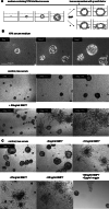
Fig. 1
Fig. 1
BMP7 induces filamentous outgrowth of spheroids in PC-3 cells but not in other prostatic cell lines. a Scheme of 3D culture method used in the study. Primarily, single cell suspension was loaded on top of extracellular matrix and allowed to form multicellular structures in 10% FBS supplemented media. When the multicellular structures reached at size of average 50 μm or more, culture media were replaced with low serum (either 0.1% or 0.5% FBS) media in presence or absence of BMP. The drawing represents morphological change of PC-3 cells in BMP-supplemented medium. b Upper panel Spheroid structures were developed from single cells of PC-3 in full serum condition. Middle and bottom panels Low serum conditions were given to cells without or with 50 ng/ml of BMP7. BMP7 stimulated morphological conversion, from spheroids to filamentous outgrowth, while cells in low serum condition maintained spheroids. Days in full serum culture and in low serum with or without BMP7 treatment were noted. c Higher concentration of BMP7 in culture medium rapidly converted cell morphology, and BMP antagonist Noggin successfully suppressed the structural change
Fig. 2
Fig. 2
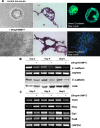
BMP7 decreases expression of E-cadherin and affects expression of transcription factors induce EMT conversion. a Upper panel shows images of PC-3 spheroids in low serum condition and bottom panel are stellate cellular filaments outgrown by BMP7. Left Panel 3D structures were shown by transmission light microscopy. Empty hollow is visible in the spheroid in the upper-left panel. Elongated fibroblastoid morphology of PC-3 cells in the filamentous structure is obvious in the bottom left panel. Scale bar, 50 μm; magnification, ×200. Middle panel Frozen sections of 3D structures were visualized by hematoxylin and eosin staining. Magnification, ×200. Right panel Immunofluorescence staining of intact 3D structure embedded in Matrigel. Loss of E-cadherin was apparent in the filamentous structure induced by 3 days of BMP7 treatment, compared with the spheroids under low serum condition. 3D structure of spheroids and filaments outgrown from spheroids were confirmed through reconstitution of Z-stack images collected by confocal microscopy (data not shown). Magnification, ×260. b Semi-quantitative PCR and Western blot analysis showed that E-cadherin expression is significantly decreased in BMP7-treated PC-3 cancer cells. Low serum condition did not have a significant effect on the level of E-cadherin. c Expression of transcription factors that mediate induction of EMT phenotype were determined by semi-quantitative PCR. Up to 4 days of BMP7 treatment, twist and slug transcription was enhanced in cancer cells. In contrast, Sip1 and Snail expressions were decreased in BMP-treated cells
Fig. 3
Fig. 3
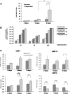
BMP7 increases invasion of PC-3 cancer cells, enhances protease activities, and upregulates protease expressions. a Number of invasive cells was counted using Matrigel invasion assays. 6 days of BMP treatment significantly enhanced cell invasiveness. *p < 0.05; **p < 0.001. b The activity of secretory proteases was evaluated using PC-3 conditioned media from cells previously cultured in low serum condition or in BMP-supplemented media for period indicated in graph legend. SensoLyte™520 MMP Substrate Sampler Kit was used. c Transcription of proteases was examined using quantitative PCR. Expression of each protease was normalized to the expression in low serum condition for 2 days of culture. 2-Delta Ct method was used to calculate mRNA change in folds and statistical assay (paired t test) was carried out to assure the statistically significant difference between BMP-treated and BMP-untreated samples. *p < 0.05; **p < 0.001
Fig. 4
Fig. 4
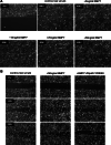
Inhibition of Akt and Erk pathways decreases cell migration of BMP-activated cells. Scratch assays were performed to compare motility of cells in low serum, with BMP7 treatment or in combination of BMP7 and various kinase inhibitors. a A 6 ng/ml or higher concentration of BMP7 stimulated cell migration compared to low serum control. b Use of LY294002, a PI3K/Akt inhibitor, counteracted the BMP7 effect on both cell migration and morphology. c Chemical inhibitor of MEK1/2, U0126 that inhibits Erk phosphorylation and activation, significantly reduced migration of cells in BMP7 culture. d Activation of signal transduction of BMP was examined by Western blot analysis. Akt phosphorylation was increased in BMP-treated cells and Erk activation was shown after 6 days of culture
Fig. 4
Fig. 4
Inhibition of Akt and Erk pathways decreases cell migration of BMP-activated cells. Scratch assays were performed to compare motility of cells in low serum, with BMP7 treatment or in combination of BMP7 and various kinase inhibitors. a A 6 ng/ml or higher concentration of BMP7 stimulated cell migration compared to low serum control. b Use of LY294002, a PI3K/Akt inhibitor, counteracted the BMP7 effect on both cell migration and morphology. c Chemical inhibitor of MEK1/2, U0126 that inhibits Erk phosphorylation and activation, significantly reduced migration of cells in BMP7 culture. d Activation of signal transduction of BMP was examined by Western blot analysis. Akt phosphorylation was increased in BMP-treated cells and Erk activation was shown after 6 days of culture
Fig. 5
Fig. 5
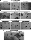
Akt and Erk pathways mediate morphological change of EMT induced by BMP7 in 3D. a Inhibition of PI3K/Akt effectively prevented the filamentous structure formation causes by BMP7, at all concentration of inhibitor used, 5 to 20 μM/ml. b Erk inhibitor was able to suppress the morphologic change at 10 μM concentration but the effect was less obvious at a lower concentration, 1 and 5 μM. c Dorsomorphin, specific chemical inhibitor of BMP-Smad signaling rather induced apoptosis in cells in 3D culture. d Both JNK inhibitor SP600125 and p38 kinase inhibitor SB203580 did not inhibit the BMP7 effect on 3D structure

"VSports最新版本" References

    1. Thiery JP, et al. Epithelial-mesenchymal transitions in development and disease. Cell. 2009;139(5):871–890. doi: 10.1016/j.cell.2009.11.007. - DOI - PubMed
    1. Weinberg RA. Twisted epithelial-mesenchymal transition blocks senescence. Nat Cell Biol. 2008;10(9):1021–1023. doi: 10.1038/ncb0908-1021. - DOI (VSports) - PubMed
    1. Polyak K, Weinberg RA. Transitions between epithelial and mesenchymal states: acquisition of malignant and stem cell traits. Nat Rev Cancer. 2009;9(4):265–273. doi: 10.1038/nrc2620. - DOI - PubMed
    1. Yang S, et al. Diverse biological effect and Smad signaling of bone morphogenetic protein 7 in prostate tumor cells. Cancer Res. 2005;65(13):5769–5777. doi: 10.1158/0008-5472.CAN-05-0289. - DOI - PubMed
    1. Rhee S, Grinnell F. Fibroblast mechanics in 3D collagen matrices. Adv Drug Deliv Rev. 2007;59(13):1299–1305. doi: 10.1016/j.addr.2007.08.006. - DOI - PMC - PubMed

Publication types

VSports最新版本 - MeSH terms