Skip to main page content
U.S. flag

An official website of the United States government

Dot gov

The . gov means it’s official. Federal government websites often end in . gov or . mil. Before sharing sensitive information, make sure you’re on a federal government site VSports app下载. .

Https

The site is secure V体育官网. The https:// ensures that you are connecting to the official website and that any information you provide is encrypted and transmitted securely. .

. 2011 Oct 10;10(10):1034-43.
doi: 10.1016/j.dnarep.2011.08.002. Epub 2011 Aug 30.

Processing of DNA structures via DNA unwinding and branch migration by the S. cerevisiae Mph1 protein (VSports app下载)

Affiliations

"V体育官网" Processing of DNA structures via DNA unwinding and branch migration by the S. cerevisiae Mph1 protein

Xiao-Feng Zheng et al. DNA Repair (Amst). .

VSports最新版本 - Abstract

The budding yeast Mph1 protein, the putative ortholog of human FANCM, possesses a 3' to 5' DNA helicase activity and is capable of disrupting the D-loop structure to suppress chromosome arm crossovers in mitotic homologous recombination. Similar to FANCM, genetic studies have implicated Mph1 in DNA replication fork repair VSports手机版. Consistent with this genetic finding, we show here that Mph1 is able to mediate replication fork reversal, and to process the Holliday junction via DNA branch migration. Moreover, Mph1 unwinds 3' and 5' DNA Flap structures that bear key features of the D-loop. These biochemical results not only provide validation for a role of Mph1 in the repair of damaged replication forks, but they also offer mechanistic insights as to its ability to efficiently disrupt the D-loop intermediate. .


Figures

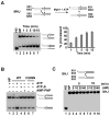
Figure 1
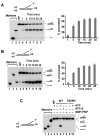
Figure 1. Processing of the 5′ Flap structure by Mph1
(A), (B) 5′ Flap substrates labeled with 32P (*) on either the short (A) or bottom strand (B) were incubated with Mph1 (2 nM) for indicated times. The results were quantified and graphed. Each experiment was performed in triplicate, and mean values are shown with standard deviation. (C) To show dependence of 5′ Flap unwinding on ATP hydrolysis, the mph1 D209N mutant (2 nM) was examined and ATP was replaced by ATP-γ-S or AMP-PNP. The incubation time was 10 min. In all the experiments, the substrate concentration was 5 nM and the reaction temperature was 30°C. Symbol: NP, minus Mph1 control.
Figure 2
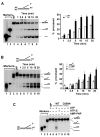
Figure 2. Processing of the 3′ Flap structure by Mph1
(A), (B) 3′ Flap substrates labeled with 32P (*) on either the short (A) or bottom strand (B) were incubated with Mph1 (2 nM) for the indicated times. The results were quantified and graphed. Each experiment was performed in triplicate, and mean values are shown with standard deviation. (C) To show dependence of 3′ Flap unwinding on ATP hydrolysis, the mph1 D209N mutant (2 nM) was examined and ATP was replaced by ATP-γ-S or AMP-PNP. The incubation time was 10 min. In all the experiments, the substrate concentration was 5 nM and the reaction temperature was 30°C. Symbol: NP, minus Mph1 control
Figure 3
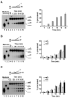
Figure 3. Relevance of the DNA branch point in the Flap substrates processing
(A) 5′ Flap substrate, (B) 3′ Flap substrate, and (C) 3′ Flap substrate containing a 10-nt 5′ tail on the short strand, all labeled with 32P (*) on the bottom strand, were incubated with Mph1 (2 nM) for the indicated times. The results were quantified and graphed. Each experiment was performed in triplicate, and mean values are shown with standard deviation. In all the experiments, the substrate concentration was 5 nM and the reaction temperature was 20°C. Symbol: NP, minus Mph1 control.
Figure 4
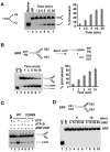
Figure 4. Replication fork regression by Mph1
(A) 32P-labeled (*) Y DNA was incubated with Mph1 (2 nM) for the indicated times. The results were quantified and graphed. Each experiment was performed in triplicate, and mean values are shown with standard deviation. (B) 32P-labeled (*) movable replication fork DNA (MRF) was incubated with Mph1 (1 nM) for the indicated times. The results were quantified and graphed. Each experiment was performed in triplicate, and mean values are shown with standard deviation. Note that the substrate harbored an A/C mismatch to minimize spontaneous regression. (C) To show dependence of replication fork regression on ATP hydrolysis, the mph1 D209N mutant (1 nM) was examined and ATP was replaced by ATP-γ-S or AMP-PNP. The incubation time was 5 min. (D) 32P-labeled static replication fork (SRF) was incubated with Mph1 (5 to 40 nM) for 5 and 10 min. In all the experiments, the substrate concentration was 5 nM and the reaction temperature was 30°C. Symbols: NP, minus Mph1 control; HD, heat-denatured substrate.
Figure 5
Figure 5. Branch migration of the Holliday junction by Mph1
(A) 32P-labeled (*) movable Holliday junction (MHJ) was incubated with Mph1 (1 nM) for the indicated times. The results were quantified and graphed. Each experiment was performed in triplicate, and mean values are shown with standard deviation. Note that the indicated positions in the two right arms of the HJ harbored a different base pair (A/T or G/C) to minimize spontaneous branch migration. (B) To show dependence of branch migration on ATP hydrolysis, the mph1 D209N mutant (1 nM) was examined and ATP was replaced by ATP-γ-S or AMP-PNP. The incubation time was 5 min. (C) 32P-labeled static Holliday junction (SHJ) was incubated with Mph1 (5 to 40 nM) for 5 and 10 min. In all the experiments, the substrate concentration was 5 nM and the reaction temperature was 30°C. Symbols: NP, minus Mph1 control; HD, heat-denatured substrate.
Figure 6
Figure 6. Comparison of Mph1, Srs2, and Sgs1 for fork regression and branch migration
32P-labeled movable replication fork (MRF) and movable Holliday junction (MHJ) were incubated with Mph1, Srs2, or Sgs1 (5 nM each) for 5 min. The results were quantified and graphed. Each experiment was performed in triplicate, and mean values are shown with standard deviation. In all the experiments, the substrate concentration was 5 nM and the reaction temperature was 30°C. Symbols: NP, no DNA motor protein added; HD, heat-denatured substrate.
Figure 7
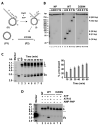
Figure 7. Fork regression and branch migration examined using a plasmid sized sigma structure
(A) Schematic representation of the 32P-labeled sigma substrate and the outcome of Mph1–mediated regression and branch migration. A, B, E, F, and N denote sites for the restriction endonucleases AvrI, BamHI, EcoRI, AflIII, and AlwNI, respectively. The formation of a regressed fork structure and branch migration of the four-way junction structure can be monitored by restriction digest. Branch migration of the four-way junction point over 2.9 kb yields a 32P-labeled linear duplex. The asterisk denotes the 32P label. (B) The sigma substrate was incubated with Mph1 or mph1 D209N (2 nM) for 20 min and then treated with the indicated restriction enzymes (see (A) for definition). (C) The sigma substrate was incubated with Mph1 (2 nM) for the indicated times and then subjected to agarose gel electrophoresis and phosphorimaging analysis.*** The results were quantified and graphed. The experiment was performed in triplicate, and mean values are shown with standard deviation. (D) To show dependence of fork regression and branch migration on ATP hydrolysis, the mph1 D209N mutant (2 nM) was examined and ATP was replaced by ATP-γ-S or AMP-PNP. The incubation time was 10 min. In all the experiments, the substrate concentration was 0.75 nM and the reaction temperature was 30°C. Symbols: S, I, P1, and P2 represent the sigma substrate, intermediates harboring the regressed fork, product 1, and product 2, respectively; NP, minus Mph1 control. In (C) and (D), free 32P-labeled pG46B plasmid used in substrate construction is denoted by**, and the species resulting from the annealing of linearized pG46B and pG68A is denoted by ***.
Figure 8
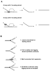
Figure 8. Functional attributes of Mph1 germane for biological functions
(A) Dissociation of D-loop structures by Mph1 in HR regulation. Our results suggest that Mph1 utilizes its structure-specific unwinding activity to disrupt D-loops that harbor a 3′ invading strand but its 3′-5′ helicase activity to dissociate D-loops that harbor a 5′ invading strand. (B) Relevance of replication fork regression and branch migration activities of Mph1 in replication fork restart. To restart the replication fork, the “chicken foot” structure in step (4) can be dissociated by reverse branch migration or used as the substrate to initiate homologous recombination. For discussions see the review by Atkinson and McGlynn [2].

References

    1. Ciccia A, Elledge SJ. The DNA damage response: making it safe to play with knives. Mol Cell. 2010;40:179–204. - PMC - PubMed
    1. Atkinson J, McGlynn P. Replication fork reversal and the maintenance of genome stability. Nucleic Acids Res. 2009;37:3475–3492. - PMC (V体育安卓版) - PubMed
    1. Ralf C, Hickson ID, Wu L. The Bloom’s syndrome helicase can promote the regression of a model replication fork. J Biol Chem. 2006;281:22839–22846. - PubMed
    1. Blastyak A, Pinter L, Unk I, Prakash L, Prakash S, Haracska L. Yeast Rad5 protein required for postreplication repair has a DNA helicase activity specific for replication fork regression. Mol Cell. 2007;28:167–175. - PMC - PubMed
    1. Blastyak A, Hajdu I, Unk I, Haracska L. Role of double-stranded DNA translocase activity of human HLTF in replication of damaged DNA. Mol Cell Biol. 2010;30:684–693. - PMC - PubMed

"V体育官网" Publication types

"V体育ios版" MeSH terms