V体育安卓版 - Skip to main page content
U.S. flag

An official website of the United States government

Dot gov

The . gov means it’s official. Federal government websites often end in . gov or VSports app下载. mil. Before sharing sensitive information, make sure you’re on a federal government site. .

Https

The site is secure. The https:// ensures that you are connecting to the official website and that any information you provide is encrypted and transmitted securely. V体育官网.

. 2011 Nov;31(11):2624-33.
doi: 10.1161/ATVBAHA.111.232827.

Paraoxonase-2 modulates stress response of endothelial cells to oxidized phospholipids and a bacterial quorum-sensing molecule

Affiliations

Paraoxonase-2 modulates stress response of endothelial cells to oxidized phospholipids and a bacterial quorum-sensing molecule

Juyong Brian Kim et al. Arterioscler Thromb Vasc Biol. 2011 Nov.

VSports在线直播 - Abstract

Objective: Chronic infection has long been postulated as a stimulus for atherogenesis. Pseudomonas aeruginosa infection has been associated with increased atherosclerosis in rats, and these bacteria produce a quorum-sensing molecule 3-oxo-dodecynoyl-homoserine lactone (3OC12-HSL) that is critical for colonization and virulence. Paraoxonase 2 (PON2) hydrolyzes 3OC12-HSL and also protects against the effects of oxidized phospholipids thought to contribute to atherosclerosis. We now report the response of human aortic endothelial cells (HAECs) to 3OC12-HSL and oxidized 1-palmitoyl-2-arachidonoyl-sn-glycero-3-phosphocholine (Ox-PAPC) in relation to PON2 expression. VSports手机版.

Methods and results: Using expression profiling and network modeling, we identified the unfolded protein response (UPR), cell cycle genes, and the mitogen-activated protein kinase signaling pathway to be heavily involved in the HAEC response to 3OC12-HSL V体育安卓版. The network also showed striking similarities to a network created based on HAEC response to Ox-PAPC, a major component of minimally modified low-density lipoprotein. HAECs in which PON2 was silenced by small interfering RNA showed increased proinflammatory response and UPR when treated with 3OC12-HSL or Ox-PAPC. .

Conclusion: 3OC12-HSL and Ox-PAPC influence similar inflammatory and UPR pathways. Quorum sensing molecules, such as 3OC12-HSL, contribute to the proatherogenic effects of chronic infection. The antiatherogenic effects of PON2 include destruction of quorum sensing molecules V体育ios版. .


Figures
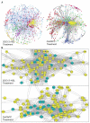
Figure 1
Figure 1. Weighted Gene Co-expression Network of HAEC transcriptome
(A) Edge-weighted spring embedded layout via Cytoscape of network created from HAEC treated with 3OC12-HSL (Left) and Ox-PAPC (Right). Nodes were colored to match the module identified from average linkage hierarchical clustering with topological overlap dissimilarity measure as described in methods. (Left) Pearson correlation with Beta=14 was used to create an adjacency matrix (ADJ) for all genes within the network and 1-ADJ was used as measure of dissimilarity. The dissimilarity index was used as the interaction strength between nodes to create a network. 925 nodes were included in total following the screen to create 12714 edges. (Right) Pearson correlation with Beta=5 was used to create ADJ within the network and dissimilarity index was used as interaction strength to create network with 827 nodes and 12000 edges; (B) Close-up view of the yellow modules from HAEC treated with 3OC12-HSL (Left) and Ox-PAPC (Right). The UPR genes within the module are highlighted in green.
Figure 2
Figure 2
PON2 knockdown resulted in more severe response of the HAEC to 3OC12-HSL treatment. (A) Determination of the ability of membrane protein isolated from control and PON2 siRNA transfected HAEC to hydrolyze 3OC12-HSL. Five μg of membrane protein from HAEC transfected with control or PON2 siRNAs was incubated with 10 μM of 3OC12-HSL for various periods of time (0 to 90 min). Afterwards, the remaining 3OC12-HSL in the reaction mixture was assayed using a bioassay. (B) and (C) Determination of the ability of intact HAEC transfected with control or PON2 siRNAs to hydrolyze 3OC12-HSL. PON2 or control siRNA transfected HAEC were incubated with 25 μM (B) or 50 μM (C) of 3OC12-HSL for 0, 2, or 4 hr before 6 μl of the media was removed and assayed for remaining 3OC12-HSL level using the bioassay. (D) Cell metabolism of PON2 or control siRNA transfected HAEC after 3OC12-HSL treatment. One day after siRNA transfection, cells were treated with various concentrations of 3OC12-HSL for 17 hrs before cell metabolism was measured. For A-D: *: p < 0.05, **: p < 0.01, ***: p < 0.0001, PON2 siRNA transfected cells vs. control siRNA transfected cells. (E) Expression of UPR genes in PON2 or control siRNA transfected HAEC with or without 3OC12-HSL treatment. Two days after siRNA transfection, cells were treated with 0 or 50 μM of 3OC12-HSL (C12) for 4 hrs before total RNA was isolated for quantitative PCR analysis. **: p < 0.01, vehicle treated-PON2 siRNA transfected cells vs. vehicle treated-control siRNA transfected cells. #: p < 0.05, ##: p < 0.01, C12 treated-PON2 siRNA transfected cells vs. C12 treated-control siRNA transfected cell. (F) Western blot analysis of UPR and apoptosis markers in PON2 or control siRNA transfected HAEC treated with 50 μM of 3OC12-HSL for various time periods (0 to 60 min, upper panel). Quantification of western blot data is shown in lower panel of (F). β actin was used as the loading control. Values for phospho-eIF2α or cleaved caspase 7 protein were normalized to β-actin. Relative protein level was calculated as (protein of interest/β-actin) of the PON2 siRNA treated sample at time X / (protein of interest/β-actin) of the control siRNA treated sample at the same time X. (G) and (I) Increased expression of calcium pathway genes (G) and inflammatory genes (I) in PON2 siRNA transfected HAEC with 3OC12-HSL treatment. For G, and I: *: p < 0.05, **: p < 0.01, ***: p < 0.0001, vehicle treated-PON2 siRNA transfected cells vs. vehicle treated-control siRNA transfected cells. #: p < 0.05, ##: p < 0.01, ###: p < 0.0001, C12 treated-PON2 siRNA transfected cells vs. C12 treated-control siRNA transfected cell. (H) Determination of NF-κB activation by Western blot analysis of phospho-p65. Cells were treated with 50 μM of 3OC12-HSL (C12) for various time periods (0 to 240 min) before total cell lysate was harvested for western blot analysis.
Figure 2
Figure 2
PON2 knockdown resulted in more severe response of the HAEC to 3OC12-HSL treatment. (A) Determination of the ability of membrane protein isolated from control and PON2 siRNA transfected HAEC to hydrolyze 3OC12-HSL. Five μg of membrane protein from HAEC transfected with control or PON2 siRNAs was incubated with 10 μM of 3OC12-HSL for various periods of time (0 to 90 min). Afterwards, the remaining 3OC12-HSL in the reaction mixture was assayed using a bioassay. (B) and (C) Determination of the ability of intact HAEC transfected with control or PON2 siRNAs to hydrolyze 3OC12-HSL. PON2 or control siRNA transfected HAEC were incubated with 25 μM (B) or 50 μM (C) of 3OC12-HSL for 0, 2, or 4 hr before 6 μl of the media was removed and assayed for remaining 3OC12-HSL level using the bioassay. (D) Cell metabolism of PON2 or control siRNA transfected HAEC after 3OC12-HSL treatment. One day after siRNA transfection, cells were treated with various concentrations of 3OC12-HSL for 17 hrs before cell metabolism was measured. For A-D: *: p < 0.05, **: p < 0.01, ***: p < 0.0001, PON2 siRNA transfected cells vs. control siRNA transfected cells. (E) Expression of UPR genes in PON2 or control siRNA transfected HAEC with or without 3OC12-HSL treatment. Two days after siRNA transfection, cells were treated with 0 or 50 μM of 3OC12-HSL (C12) for 4 hrs before total RNA was isolated for quantitative PCR analysis. **: p < 0.01, vehicle treated-PON2 siRNA transfected cells vs. vehicle treated-control siRNA transfected cells. #: p < 0.05, ##: p < 0.01, C12 treated-PON2 siRNA transfected cells vs. C12 treated-control siRNA transfected cell. (F) Western blot analysis of UPR and apoptosis markers in PON2 or control siRNA transfected HAEC treated with 50 μM of 3OC12-HSL for various time periods (0 to 60 min, upper panel). Quantification of western blot data is shown in lower panel of (F). β actin was used as the loading control. Values for phospho-eIF2α or cleaved caspase 7 protein were normalized to β-actin. Relative protein level was calculated as (protein of interest/β-actin) of the PON2 siRNA treated sample at time X / (protein of interest/β-actin) of the control siRNA treated sample at the same time X. (G) and (I) Increased expression of calcium pathway genes (G) and inflammatory genes (I) in PON2 siRNA transfected HAEC with 3OC12-HSL treatment. For G, and I: *: p < 0.05, **: p < 0.01, ***: p < 0.0001, vehicle treated-PON2 siRNA transfected cells vs. vehicle treated-control siRNA transfected cells. #: p < 0.05, ##: p < 0.01, ###: p < 0.0001, C12 treated-PON2 siRNA transfected cells vs. C12 treated-control siRNA transfected cell. (H) Determination of NF-κB activation by Western blot analysis of phospho-p65. Cells were treated with 50 μM of 3OC12-HSL (C12) for various time periods (0 to 240 min) before total cell lysate was harvested for western blot analysis.
Figure 3
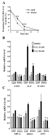
Figure 3. PON2 overexpression decreased 3OC12-HSL induced inflammatory and UPR response in HeLa cells
(A) HeLa cells transfected with a GFP or a human PON2 expression vector were incubated with 50 μM of 3OC12-HSL for 0 to 2 hr before 6 μl of the media was removed and assayed for remaining 3OC12-HSL level using the bioassay. **: p < 0.01, ***: p < 0.0001, GFP vs. PON2 transfected cells. (B), (C) Decreased expression of inflammatory genes (B) and UPR genes (C) in 3OC12-HSL treated HeLa cells transfected with a human PON2 expression vector. Two concentrations of 3OC12-HSL (C12) were used: 25 μM and 50 μM. Treatment time was 4 hr. *: p < 0.05, GFP- vs. PON2-transfected cells receiving the same treatment.
Figure 4
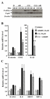
Figure 4. EP2 is involved in 3OC12-HSL induced NF-kB activation
(A) Western blot analysis of HAEC treated with vehicle control, DMSO (D), 10 μM of AH6809 (AH), 50 μM of 3OC12-HSL (C12), or 10 μM of AH6809 and 50 μM of 3OC12-HSL (C12 + AH). (B) and (C) gene expression analysis of HAEC after 4 hr treatment of the same agents as described in (A). *: p < 0.05, **: p < 0.01, C12 treated vs. C12 + AH6809 treated.
Figure 5
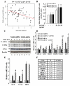
Figure 5. PON2 protects against Ox-PAPC induced oxidative stress and UPR response in HAEC
(A) Inverse correlation between PON2 mRNA levels and intracellular superoxide levels (as determined by the NBT reduction assay) among 96 primary cultures of HAEC. (B) Increased intracellular superoxide levels in PON2 siRNA transfected HAEC with or without Ox-PAPC treatment. Control or PON2 siRNAs transfected HAEC were treated with 0, or 50 μg /ml of Ox-PAPC for 1 hr before intracellular superoxide levels were measured. *: p < 0.05, PON2 siRNA transfected cells vs. control siRNA transfected cells. (C) Western blot analysis of UPR markers of HAEC transfected with PON2 or control siRNA and treated with 50 μg /ml of Ox-PAPC for 0 to 4 hrs. (D) Induction of UPR genes in HAEC transfected with PON2 or control siRNA and treated with 0 or 50 μg /ml of Ox-PAPC for 4 hrs. (E) Induction of expression of IL-8, CCL2, and HO-1 genes in HAEC transfected with PON2 or control siRNA and treated with 0 or 50 μg /ml of Ox-PAPC for 4 hrs. #: p < 0.05, ##: p < 0.01, Ox-PAPC treated-PON2 siRNA transfected cells vs. Ox-PAPC treated-control siRNA transfected cell. (F) Correlation between basal PON2 mRNA level and fold change of UPR genes upon Ox-PAPC treatment among 96 primary cultures of HAEC. Expression data were obtained from our previous publication. Briefly, HAEC from each donor were treated in duplicate with media (basal level) or media + 40 μg/ml Ox-PAPC (Ox-PAPC) for 4 hr before RNA isolation. RNA expression level was determined by Affymetrix HT-HU133A Microarrays. The fold change of Ox-PAPC over basal level of UPR gene of interest, which was calculated as the (log2(Ox-PAPC) − log2(basal)), was correlated with basal level of PON2 mRNA.

"V体育ios版" References

    1. Stassen FR, Vainas T, Bruggeman CA. Infection and atherosclerosis. An alternative view on an outdated hypothesis. Pharmacol Rep. 2008;60:85–92. - VSports app下载 - PubMed
    1. Danesh J. Antibiotics in the prevention of heart attacks. Lancet. 2005;365:365–367. - PubMed
    1. Leinonen M, Saikku P. Evidence for infectious agents in cardiovascular disease and atherosclerosis. Lancet Infect Dis. 2002;2:11–17. - PubMed
    1. Libby P, Egan D, Skarlatos S. Roles of infectious agents in atherosclerosis and restenosis: An assessment of the evidence and need for future research. Circulation. 1997;96:4095–4103. - PubMed
    1. Turkay C, Saba R, Sahin N, Altunbas H, Ozbudak O, Akkaya B, Ozbilim G, Colbasi I, Turkay M, Ogunc D, Bayezid O. Effect of chronic pseudomonas aeruginosa infection on the development of atherosclerosis in a rat model. Clin Microbiol Infect. 2004;10:705–708. - "V体育ios版" PubMed

Publication types

"V体育ios版" MeSH terms