Skip to main page content
U.S. flag

An official website of the United States government

Dot gov

The VSports app下载. gov means it’s official. Federal government websites often end in . gov or . mil. Before sharing sensitive information, make sure you’re on a federal government site. .

Https

The site is secure V体育官网. The https:// ensures that you are connecting to the official website and that any information you provide is encrypted and transmitted securely. .

. 2011;6(7):e21889.
doi: 10.1371/journal.pone.0021889. Epub 2011 Jul 18.

Interaction between the triglyceride lipase ATGL and the Arf1 activator GBF1

Affiliations

Interaction between the triglyceride lipase ATGL and the Arf1 activator GBF1

Emy Njoh Ellong et al. PLoS One. 2011.

"VSports app下载" Abstract

The Arf1 exchange factor GBF1 (Golgi Brefeldin A resistance factor 1) and its effector COPI are required for delivery of ATGL (adipose triglyceride lipase) to lipid droplets (LDs). Using yeast two hybrid, co-immunoprecipitation in mammalian cells and direct protein binding approaches, we report here that GBF1 and ATGL interact directly and in cells, through multiple contact sites on each protein. The C-terminal region of ATGL interacts with N-terminal domains of GBF1, including the catalytic Sec7 domain, but not with full-length GBF1 or its entire N-terminus. The N-terminal lipase domain of ATGL (called the patatin domain) interacts with two C-terminal domains of GBF1, HDS (Homology downstream of Sec7) 1 and HDS2 VSports手机版. These two domains of GBF1 localize to lipid droplets when expressed alone in cells, but not to the Golgi, unlike the full-length GBF1 protein, which localizes to both. We suggest that interaction of GBF1 with ATGL may be involved in the membrane trafficking pathway mediated by GBF1, Arf1 and COPI that contributes to the localization of ATGL to lipid droplets. .


Conflict of interest statement

Competing Interests: The authors have declared that no competing interests exist.

Figures

Figure 1
Figure 1. ATGL interacts with Arf1 GEF catalytic Sec7 domains.
A- Yeast strain AH109 was transformed with the pACT2 library plasmid containing amino acids 367–504 of ATGL fused to the activation domain of Gal4p, and pGBKT7 vectors carrying the indicated Sec7 domain fused to the DNA-binding domain of Gal4p. Cells were spotted on the indicated selective plates and incubated for three days at 30°C. B-C- HA-tagged H. sapiens ATGL was coexpressed in Cos7 cells either with Venus alone, or with the indicated Venus-tagged Sec7 domain; Sec7-EK indicates a mutation of the catalytic glutamate residue to lysine. Immunoprecipitation was carried out with anti-GFP antibodies, and Cos7 cell lysates or eluted proteins after immunoprecipitation were analyzed by Western blotting using anti-HA antibody (left panels). Lysates from cells expressing the indicated Venus-GBF1 construct were analyzed by Western blotting using anti-GFP antibody (right panels). B- Wild type and E794K mutant Sec7 domains of GBF1. C- EK mutant Sec7 domains of GBF1, BIG1, BIG2, ARNO and EFA6.
Figure 2
Figure 2. Fragments of Homo sapiens GBF1 and ATGL proteins used in this study.
A- Schematic diagram of GBF1 showing domains, with regions used in the interaction experiments shown below. Amino acid numbers for the beginning and end of each fragment are indicated. Numbers in black are for GBF1, numbers in red refer to BIG1. B- Schematic diagram of ATGL, with regions used in interaction experiments shown below. The constructs indicated in bold are those that were demonstrated to interact directly when expressed in E. coli and were also shown to interact by co-immunoprecipitation and yeast two-hybrid methods (with the exception of the HDS1-ATGL patatin domain interaction which was not detected in yeast two-hybrid); see Table 2 for details. Note that slight variations in the borders of GBF1 constructs were used for the yeast two-hybrid experiments (see Table 1), but essentially the same domains were used for all interaction studies.
Figure 3
Figure 3. Yeast two-hybrid interactions between ATGL regions and GBF1 Sec7 and DCB domains.
Yeast strain AH109 was transformed with the indicated GBF1 domains in pGADT7, along with the indicated regions of ATGL in pGBKT7 plasmids. 10-fold serial dilutions of each doubly transformed strain were spotted onto the indicated plates and incubated for 2 days at 30°C. A- GBF1 Sec7 domain. B- GBF1 DCB domain.
Figure 4
Figure 4. Interactions between GBF1 and ATGL assayed by co-immunoprecipitation.
A- Immmunoprecipitation (IP) of endogenous proteins. In the left panel, ATGL antibody (GFP antibody for the negative control) was used for the IP, and the western blot was performed with GBF1 antibody. In the right panel, GBF1 antibody (GFP antibody for negative control) was used for the IP, and the western blot was performd with ATGL antibody. B- HA-tagged H. sapiens ATGL (either full-length or C-terminally truncated, as indicated) was coexpressed in Cos7 cells either with Venus alone, or with the indicated Venus-tagged GBF1 region. Immunoprecipitation was carried out with anti-GFP antibodies, and Cos7 cell lysates or eluted proteins after immunoprecipitation were analyzed by Western blotting using anti-HA antibody. For Western blots of the GFP-immunoprecipitated bait proteins, see Figure S3A. C- Quantification represents the level of ATGL in the indicated immunoprecipitate, after subtracting the value for pVenus and normalizing to the fragment with the highest level of interaction, that of the ΔDCB construct. This value was set to 100% for each ATGL construct; as shown in part B, these values were equivalent for full-length ATGL and ATGL(1–366). Mean and standard deviation of 2–4 independent experiments are shown. D- Co-immunoprecipitations from Cos7 cells expressing the indicated portion of HA-tagged H. sapiens ATGL and Venus-tagged GBF1 C-terminal regions were carried out as in part B. E- Quantifications were carried out as in part C, with values normalized to ATGL levels in HDS1 immunoprecipitations. Mean and standard deviation of 2–4 independent experiments are shown. Quantifications were carried out only for the three GBF1 fragments whose expression levels were approximately equivalent; HDS2 was expressed at a significantly higher level (see Figure S1A).
Figure 5
Figure 5. Catalytic activity of ATGL is not required for LD localization or interaction with GBF1.
A- Immuno-EM of HeLa cells expressing GFP-tagged H. sapiens ATGL-S47A were treated with 200 µM oleic acid for 15 hours and labeled with gold-conjugated antibodies against GFP. The majority of gold particles (examples indicated by arrows) are found on the surface of LDs. LD, lipid droplet. B- Co-immunoprecipitations from Cos7 cells expressing HA-tagged H. sapiens ATGL-S47A or ATGL(1–366)-S47A and Venus-tagged GBF1 fragments were carried out as in Figures 1 and 4; Western blot of immunoprecipitates probed with anti-HA antibodies (upper panel); Cos7 cell extracts blotted with anti-GFP antibodies (lower panel).
Figure 6
Figure 6. Direct interactions between domains of GBF1 and ATGL.
A- Purified GST, GST-tagged ATGL(1–366) or GST-tagged ATGL(300–504) was bound to glutathione Sepharose beads, then incubated with E. coli lysates expressing the His-tagged DCB domain of GBF1. Eluted proteins were analyzed by Western blotting using His antibody. B- and C- The indicated His-tagged GBF1 domain expressed in E. coli was bound to Ni Sepharose 6 Fast Flow, then purified GST or the indicated GST-tagged domain of ATGL were incubated with the protein-bound beads. Eluted proteins were analyzed by Western blotting using GST antibody. B- The HDS1-His domain of GBF1 bound to Ni Sepharose beads. C- The His-tagged Sec7 domain of GBF1 bound to Ni Sepharose beads.
Figure 7
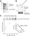
Figure 7. The GBF1 Sec7 domain directly interacts with the ATGL (300–504) domain in vitro.
A- Coomassie stained gel showing purified recombinant GST-tagged ATGL (300–504) (1 µg, lane 1) and His-Sec7-GBF1(710–894) (1 µg, lane 2). M, molecular weight markers. B- 0.1 nmole of His-Sec7-GBF1(710–894) were immobilized on a Ni Sepharose 6 Fast Flow resin, then incubated with either GST or GST-tagged ATGL(300–504) (0.5 µM each) for 2.5 hours at 4°C. After washes, proteins were eluted, separated by SDS-PAGE followed by immunoblotting with anti-GST antibody (upper panel) or by staining with Coomassie Brilliant Blue (lower panel). GST-tagged ATGL(300-504) specifically bound to the His-Sec7-GBF1 fragment. C- Quantitative analysis of the interaction between His-tagged GBF1 Sec7 domain and GST-tagged ATGL (300–504) was performed. Various concentrations (1–20 µM) of GST-tagged ATGL (300–504) were added to immobilized His-Sec7-GBF1(710–894) domain (0.1 nmole). Each sample was washed and subjected to SDS-PAGE followed by immunoblotting with anti-GST antibody (upper panel). The amount of bound versus free GST-tagged ATGL (300–504) domain is plotted (lower panel). Scatchard analysis indicated an apparent Kd of 2.5±0.3 µM (inset).
Figure 8
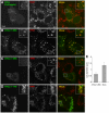
Figure 8. The GBF1 HDS1 and HDS2 domains localize to lipid droplets in cells.
HeLa cells transfected with Venus-tagged HDS1 and/or HDS2 domains of GBF1 as indicated were treated with 400 µM oleic acid for 3 hours, then immunostained with antibodies against TIP47 (middle panels). Venus fluorescence is shown on the left; the merge image on the right. Bar: 10 µm. A- Cells expressing the region of GBF1 from the beginning of HDS1 to the end of HDS2, N-terminally tagged with Venus. B- Cells expressing the Venus-tagged HDS1 domain. C- Cells expressing Venus-HDS2. Bar, 10 µm.
Figure 9
Figure 9. The C-terminal region of ATGL regulates its localization to LDs in cells.
HeLa cells transfected with the indicated ATGL constructs tagged at their C-terminus with GFP were treated with 400 µM OA for 3 hours, then immunostained with antibodies against TIP47 (middle panel). GFP fluorescence is shown on the left; the merge image on the right. Bar: 10 µm. A- Cells expressing full-length ATGL(1–504)-GFP. B- Cells expressing ATGL(1–366)-GFP. C- Cells expressing ATGL(1–289)-GFP. D- Cells expressing ATGL(1–178)-GFP. E- LD diameters were measured in cells expressing ATGL(1–289)-GFP and in control cells. Mean and standard deviation of three independent experiments are shown. Bar, 10 µm.

"VSports" References

    1. Bonifacino JS, Glick BS. The mechanisms of vesicle budding and fusion. Cell. 2004;116:153–166. - PubMed
    1. Lee MC, Miller EA, Goldberg J, Orci L, Schekman R. Bi-directional protein transport between the ER and Golgi. Annu Rev Cell Dev Biol. 2004;20:87–123. - "VSports手机版" PubMed
    1. Gillingham AK, Munro S. The small G proteins of the Arf family and their regulators. Annu Rev Cell Dev Biol. 2007;23:579–611. - PubMed
    1. Manolea F, Claude A, Chun J, Rosas J, Melancon P. Distinct functions for Arf guanine nucleotide exchange factors at the Golgi complex: GBF1 and BIGs are required for assembly and maintenance of the Golgi stack and trans-Golgi network, respectively. Mol Biol Cell. 2008;19:523–535. - PMC - PubMed
    1. Deng Y, Golinelli-Cohen MP, Smirnova E, Jackson CL. A COPI coat subunit interacts directly with an early-Golgi localized Arf exchange factor. EMBO Rep. 2009;10:58–64. - VSports app下载 - PMC - PubMed

"V体育平台登录" Publication types

MeSH terms

LinkOut - more resources