"VSports手机版" Skip to main page content
U.S. flag

An official website of the United States government

Dot gov

The . gov means it’s official. Federal government websites often end in . gov or . mil. Before sharing sensitive information, make sure you’re on a federal government site. VSports app下载.

Https

The site is secure. The https:// ensures that you are connecting to the official website and that any information you provide is encrypted and transmitted securely V体育官网. .

. 2011 Sep 9;286(36):31180-93.
doi: 10.1074/jbc.M111.258038. Epub 2011 Jul 22.

Roles of the four DNA polymerases of the crenarchaeon Sulfolobus solfataricus and accessory proteins in DNA replication

Affiliations

Roles of the four DNA polymerases of the crenarchaeon Sulfolobus solfataricus and accessory proteins in DNA replication

Jeong-Yun Choi et al. J Biol Chem. .

Erratum in

  • J Biol Chem. 2011 Nov 11;286(45):39674

VSports手机版 - Abstract

The hyperthermophilic crenarchaeon Sulfolobus solfataricus P2 encodes three B-family DNA polymerase genes, B1 (Dpo1), B2 (Dpo2), and B3 (Dpo3), and one Y-family DNA polymerase gene, Dpo4, which are related to eukaryotic counterparts. Both mRNAs and proteins of all four DNA polymerases were constitutively expressed in all growth phases. Dpo2 and Dpo3 possessed very low DNA polymerase and 3' to 5' exonuclease activities in vitro. Steady-state kinetic efficiencies (k(cat)/K(m)) for correct nucleotide insertion by Dpo2 and Dpo3 were several orders of magnitude less than Dpo1 and Dpo4 VSports手机版. Both the accessory proteins proliferating cell nuclear antigen and the clamp loader replication factor C facilitated DNA synthesis with Dpo3, as with Dpo1 and Dpo4, but very weakly with Dpo2. DNA synthesis by Dpo2 and Dpo3 was remarkably decreased by single-stranded binding protein, in contrast to Dpo1 and Dpo4. DNA synthesis in the presence of proliferating cell nuclear antigen, replication factor C, and single-stranded binding protein was most processive with Dpo1, whereas DNA lesion bypass was most effective with Dpo4. Both Dpo2 and Dpo3, but not Dpo1, bypassed hypoxanthine and 8-oxoguanine. Dpo2 and Dpo3 bypassed uracil and cis-syn cyclobutane thymine dimer, respectively. High concentrations of Dpo2 or Dpo3 did not attenuate DNA synthesis by Dpo1 or Dpo4. We conclude that Dpo2 and Dpo3 are much less functional and more thermolabile than Dpo1 and Dpo4 in vitro but have bypass activities across hypoxanthine, 8-oxoguanine, and either uracil or cis-syn cyclobutane thymine dimer, suggesting their catalytically limited roles in translesion DNA synthesis past deaminated, oxidized base lesions and/or UV-induced damage. .

V体育官网入口 - PubMed Disclaimer

VSports - Figures

FIGURE 1.
FIGURE 1.
Purified DNA polymerases and accessory proteins of S. solfataricus. The purified recombinant proteins (∼7 μg each) were separated by SDS-polyacrylamide gel electrophoresis (4–20% gradient gel, w/v) and stained with Coomassie Brilliant Blue R-250 (22, 23). The two bands for RFC are the high and low Mr subunits.
FIGURE 2.
FIGURE 2.
Growth curve of S. solfataricus strain P2 at 80 °C.
FIGURE 3.
FIGURE 3.
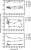
RT-PCR analyses of S. solfataricus strain P2 RNA levels as a function of growth time. RT-PCR products were separated on 2% agarose (w/v) gels and visualized by ethidium bromide staining (supplemental Fig. S1). The images were analyzed with Quantity One software (Bio-Rad). A, Dpo1, Dpo2, Dpo3, and Dpo4; B, PCNA1, PCNA2, and PCNA3; C, RFC1, RFC2, SSB, and RadA.
FIGURE 4.
FIGURE 4.
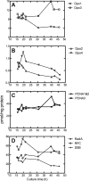
Quantitative immunoblot analyses of proteins in S. solfataricus strain P2 as a function of growth time. Images were analyzed on an Odyssey Infrared Imaging System (LI-COR) as described under “Experimental Procedures.” A, Dpo1 and Dpo2; B, Dpo3 and Dpo4; C, PCNA1/PCNA2 (these were not distinguished and are presented as the sum) and PCNA3; C, RFC, SSB, and RadA.
FIGURE 5.
FIGURE 5.
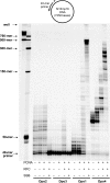
DNA polymerase (Pol) activities of Dpo1, Dpo2, Dpo3, and Dpo4. Reactions were done for 30 min at 37 °C with increasing concentrations of Dpo1 (0–20 nm), Dpo2 (0–4 μm), Dpo3 (0–4 μm), or Dpo4 (0–20 nm) with 50 nm 21-mer primer/36-G-mer template DNA as indicated (Table 1). 32P-Labeled 21-mer primer was extended in the presence of 10 mm MgCl2 and all four dNTPs (100 μm each). The reaction products were analyzed by 16% (w/v) denaturing polyacrylamide gel electrophoresis with subsequent phosphorimaging analysis.
FIGURE 6.
FIGURE 6.
DNA synthesis activities on recessed, gapped, and forked DNA substrates by Dpo1, Dpo2, Dpo3, and Dpo4 in the presence of Mg2+ or Mn2+. A, scheme of the three types of DNA substrates used. The recessed primer-template DNA was made by annealing the 32P-end-labeled primer (18-mer) and the unmodified 36-mer templates. The single-nucleotide gapped DNA was made by annealing the 32P-end-labeled primer (18-mer), the 17-mer with a 5′-phosphate (17-p-mer), and the unmodified 36-mer templates. The forked DNA was made by annealing the 32P-end-labeled primer (18-mer), the half-complementary 36-f-mer, and the unmodified 36-mer templates. All oligonucleotide sequences are listed in Table 1. *, 32P label; p, phosphate group. B, either 0.5 nm Dpo1, 150 nm Dpo2, 1 μm Dpo3, or 0.1 nm Dpo4 was incubated for 15 min at 50 °C with 50 nm DNA substrate as indicated. All 32P-labeled primers were extended with all four dNTPs (100 μm each) in the presence of 1 mm MgCl2 or MnCl2. The reaction products were analyzed by 16% (w/v) denaturing polyacrylamide gel electrophoresis with subsequent phosphorimaging analysis. r, recessed primer-template DNA; g, gapped DNA; f, forked DNA.
FIGURE 7.
FIGURE 7.
3′ → 5′ exonuclease activities of Dpo1, Dpo2, Dpo3, and Dpo4. The primer (21-mer) was annealed with unmodified 36-G-mer template (Table 1). Reactions were done for 30 min at 37 °C, with increasing concentrations of Dpo1 (0–20 nm), Dpo2 (0–4 μm), Dpo3 (0–4 μm), or Dpo4 (0–20 nm) with 50 nm primer-template DNA as indicated. 32P-labeled 21-mer primer was incubated in the presence of MgCl2 but no dNTPs at 37 °C. The reaction products were analyzed by 16% (w/v) denaturing polyacrylamide gel electrophoresis with subsequent phosphorimaging analysis. Pol, polymerase.
FIGURE 8.
FIGURE 8.
Comparison of relative DNA binding affinities of Dpo1, Dpo2, Dpo3, and Dpo4 to 24-mer·36-mer DNA by electrophoretic mobility shift assay. A, Dpo1; B, Dpo2; C, Dpo3; D, Dpo4. Reaction mixtures containing 10 nm 32P-labeled 24-mer·36-mer primer-template duplex DNA were incubated with increasing concentrations (as indicated in the figure) of each polymerase (Pol) and resolved on a 3.5% (w/v) non-denaturing polyacrylamide gel to separate the free DNA and the polymerase-DNA complex.
FIGURE 9.
FIGURE 9.
Effect of temperature on activities of nucleotide incorporation opposite G by Dpo1, Dpo2, Dpo3, and Dpo4. Reactions were done for 15 min with Dpo1 (5 nm, □), Dpo2 (0.5 μm, △), Dpo3 (1 μm, ▽), or Dpo4 (1 nm, ○) with 50 nm 21-mer primer·36-G-mer template DNA (Table 1) in the presence of 100 μm dCTP at various temperatures. The reaction products were analyzed by 16% (w/v) denaturing polyacrylamide gel electrophoresis with subsequent phosphorimaging analysis. Activities are indicated relative to the highest activity (taken as 100%) of each individual enzyme.
FIGURE 10.
FIGURE 10.
Thermostabilties of nucleotide incorporation activities of Dpo1, Dpo2, Dpo3, and Dpo4 at 60 °C. Dpo1 (24 nm, □), Dpo2 (2 μm, △), Dpo3 (6 μm, ▽), or Dpo4 (4 nm, ○) was preincubated without DNA, dNTP, and BSA for various time intervals (0–40 min) at 60 °C. Thereafter, reactions were done with the addition of the missing components (50 nm 21-mer·36-G-mer DNA, 100 μm dCTP, 100 μg/ml BSA, and 10 mm MgCl2) for 15 min at 50 °C. The reaction products were analyzed by 16% (w/v) denaturing polyacrylamide gel electrophoresis with subsequent phosphorimaging analysis. Activities are indicated relative to the activity (taken as 100%) of enzyme without preincubation.
FIGURE 11.
FIGURE 11.
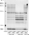
Effects of PCNA, RFC, and SSB on DNA polymerization by Dpo1, Dpo2, Dpo3, and Dpo4. Dpo1 (5 nm), Dpo2 (1 μm), Dpo3 (1 μm), or Dpo4 (5 nm) was incubated with 5 nm 40-mer primer·M13mp18 template DNA in the presence of 10 mm MgCl2, 500 μm ATP, and all four dNTPs (100 μm each) for 10 min at 50 °C, with 0.6 μm PCNA, 0.6 μm RFC, or 6 μm SSB added as indicated. The reaction products were analyzed by 8% (w/v) denaturing polyacrylamide gel electrophoresis with subsequent phosphorimaging analysis.
FIGURE 12.
FIGURE 12.
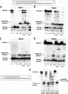
Translesion DNA synthesis across various DNA adducts by Dpo1, Dpo2, Dpo3, and Dpo4. A–D, the primer (21-mer) was annealed with each of the eight different 36-mer templates (Table 1) containing an unmodified G or hypoxanthine (H), 8-oxoG (OG), AP site, N2-MeG, O6-MeG, N2-BzG, or O6-BzG placed at the 25th position from the 3′-end. Either 20 nm Dpo1 (A), 4 μm Dpo2 (B), 4 μm Dpo3 (C), or 20 nm Dpo4 (D) was incubated for 30 min at 37 °C with 50 nm 21-mer primer·36-mer template DNA as indicated. E, the primer (17-mer) was annealed with 25-mer templates (Table 1) containing an unmodified TT or CTD placed at the 18th position from the 3′-end. Dpo1 (20 nm), Dpo2 (4 μm), Dpo3 (4 μm), or Dpo4 (20 nm) was incubated for 30 min at 37 °C with 50 nm 17-mer primer·25-mer template DNA, as indicated. All 32P-labeled primers were extended in the presence of 10 mm MgCl2 and all four dNTPs (100 μm each). The reaction products were analyzed by 16% (w/v) denaturing gel electrophoresis with subsequent phosphorimaging analysis.
FIGURE 13.
FIGURE 13.
Translesion DNA synthesis across uracil and hypoxanthine residues by Dpo1, Dpo2, Dpo3, and Dpo4. The 32P-end-labeled primer (18-mer) was annealed with each of the three different 36-mer templates (Table 1) containing an unmodified G, U, or hypoxanthine (H) at the position seven bases ahead of the primer-template junction. Either 20 nm Dpo1 (A), 4 μm Dpo2 (B), 4 μm Dpo3 (C), or 20 nm Dpo4 (D) was incubated for 30 min at 37 °C with 50 nm 21-mer primer·36-mer template DNA as indicated. All 32P-labeled primers were extended in the presence of 10 mm MgCl2 and all four dNTPs (100 μm each). The reaction products were analyzed by 16% (w/v) denaturing polyacrylamide gel electrophoresis with subsequent phosphorimaging analysis.
FIGURE 14.
FIGURE 14.
Effects of Dpo2, Dpo3, and Dpo4 on DNA polymerase (Pol) activities of Dpo1 and Dpo4. Dpo1 (1 nm) or Dpo4 (1 nm) was incubated with 5 nm 40-mer primer·M13mp18 template DNA in the presence of 10 mm MgCl2, 500 μm ATP, all four dNTPs (100 μm each), 0.6 μm PCNA, 0.6 μm RFC, and 6 μm SSB for 10 min at 50 °C, where Dpo2 (0–0.9 μm), Dpo3 (0–0.9 μm), or Dpo4 (0–10 nm) was added as indicated. The reaction products were analyzed by 8% (w/v) denaturing polyacrylamide gel electrophoresis with subsequent phosphorimaging analysis.

References

    1. Kornberg A., Baker T. A. (1992) DNA Replication, 2nd Ed., W.H. Freeman, New York
    1. Friedberg E. C., Walker G. C., Siede W., Wood R. D., Schultz R. A., Ellenberger T. (2006) DNA Repair and Mutagenesis, 2nd Ed., American Society for Microbiology Press, Washington, D. C
    1. Rothwell P. J., Waksman G. (2005) Adv. Protein Chem. 71, 401–440 - PubMed
    1. Hubscher U., Maga G., Spadari S. (2002) Annu. Rev. Biochem. 71, 133–163 - VSports注册入口 - PubMed
    1. Rivera M. C., Lake J. A. (2004) Nature 431, 152–155 - PubMed

Publication types

MeSH terms

Substances (VSports最新版本)