<u dropzone="FFvxQKT"></u> <noscript lang="fuICr2i"></noscript><ins id="s9YtfY"></ins> Skip to main page content
U.S. flag

An official website of the United States government

Dot gov

The VSports app下载. gov means it’s official. Federal government websites often end in . gov or . mil. Before sharing sensitive information, make sure you’re on a federal government site. .

Https

The site is secure. The https:// ensures that you are connecting to the official website and that any information you provide is encrypted and transmitted securely. V体育官网.

. 2011 Feb;121(2):784-99.
doi: 10.1172/JCI43757. Epub 2011 Jan 25.

Human tumors instigate granulin-expressing hematopoietic cells that promote malignancy by activating stromal fibroblasts in mice

Affiliations

Human tumors instigate granulin-expressing hematopoietic cells that promote malignancy by activating stromal fibroblasts in mice

Moshe Elkabets et al. J Clin Invest. 2011 Feb.

Abstract (V体育官网)

Systemic instigation is a process by which endocrine signals sent from certain tumors (instigators) stimulate BM cells (BMCs), which are mobilized into the circulation and subsequently foster the growth of otherwise indolent carcinoma cells (responders) residing at distant anatomical sites. The identity of the BMCs and their specific contribution or contributions to responder tumor growth have been elusive. Here, we have demonstrated that Sca1+ cKit- hematopoietic BMCs of mouse hosts bearing instigating tumors promote the growth of responding tumors that form with a myofibroblast-rich, desmoplastic stroma. Such stroma is almost always observed in malignant human adenocarcinomas and is an indicator of poor prognosis. We then identified granulin (GRN) as the most upregulated gene in instigating Sca1+ cKit- BMCs relative to counterpart control cells. The GRN+ BMCs that were recruited to the responding tumors induced resident tissue fibroblasts to express genes that promoted malignant tumor progression; indeed, treatment with recombinant GRN alone was sufficient to promote desmoplastic responding tumor growth. Further, analysis of tumor tissues from a cohort of breast cancer patients revealed that high GRN expression correlated with the most aggressive triple-negative, basal-like tumor subtype and reduced patient survival VSports手机版. Our data suggest that GRN and the unique hematopoietic BMCs that produce it might serve as novel therapeutic targets. .


Figures

Figure 1
Figure 1. Systemic instigation of responding tumor growth and stromal desmoplasia.
(A) Systemic instigation injection scheme. HMLER-HR transformed cells (responders) are injected subcutaneously into 1 flank of host mice. The opposite flank is injected with either a Matrigel control or aggressively growing tumor cell lines (instigators or noninstigators). (B) Growth kinetics of instigating and responding tumors. Of the responder cell injections, 1 of 5 formed tumors opposite Matrigel, 4 of 4 formed growing tumors opposite instigating BPLER tumors, and 0 of 5 formed tumors opposite noninstigating PC3 tumors. (C) Histopathology of resulting BPLER and HMLER-HR tumors 90 days after injection. Top panel shows αSMA staining of myofibroblasts and pericytes (brown) and hematoxylin counterstaining of nuclei (blue). Bottom panel shows Masson’s trichrome staining for collagen (blue) and nuclei counterstaining (dark pink). Scale bar: 100 μm. (D) Staining for the SV40 LgT (brown) to identify tumor cells in the resulting tumor tissues. Scale bar: 100 μm. (E and F) CellProfiler quantification of area occupied by αSMA+ staining (E) and number of LgT+ cells (F) in images of resulting tumor tissues under indicated conditions. An average of 10 images of instigating tumors and the contralateral responding tumors (Resp opp instigator) and 5 images of the responding tumor recovered opposite Matrigel (Resp opp Matrigel) were used for quantification. Data are expressed as mean ± SEM.
Figure 2
Figure 2. BMCs from instigator-bearing animals phenocopy systemic instigation.
(A) Experimental scheme to test BMC tumor supportive function: admixtures of BMCs and responding tumor cells are injected subcutaneously into host nude mice. (B) Average mass of resulting tumors 12 weeks after implantation of various indicated mixtures. Tumor incidence is indicated above bars (2 separate experiments, n = 16 per group). Data are expressed as mean ± SEM. (C) Histopathology of resulting responding tumors harvested 12 weeks after implantation of indicated mixtures. Photomicrographs show staining for αSMA (brown) and nuclei counterstained with hematoxylin (blue). Scale bar: 100 μm. (D) Experimental scheme for injecting tumor cells subcutaneously into mice that had previously been engrafted with GFP+ BMCs. (E) Merged immunofluorescent images of responding tumors that had grown for 12 weeks opposite BPLER (top) or MDA-MB-231 (bottom) instigating tumors in GFP+ BMC transplanted mice. Images represent GFP+ BM–derived cells (green); αSMA+ tumor myofibroblasts and pericytes (red); and cell nuclei (DAPI; blue). Yellow signal represents an overlap of 2 different cells, as confirmed by confocal microscopy. Scale bar: 25 μm.
Figure 3
Figure 3. Sca1+cKit hematopoietic BMCs are activated by instigating tumors.
(A) FACS density plots representing the collection of the following BMC populations from instigating tumor-bearing mice: Sca1+cKit+ (population i), Sca1 depleted (population ii), and Sca1+cKit (population iii). BMC population collected from control mice: Sca1+cKit (population iv). (B) Relative average mass of resulting tumors 12 weeks after subcutaneous injection of responder-BMC mixtures. Tumor mass was normalized to the mass of control responder tumors that had grown on their own (3 separate experiments; n = 18 per group). (C) Histopathology of responding tumors resulting from the indicated admixtures. Top panel shows αSMA staining of myofibroblasts and pericytes (brown) and hematoxylin counterstaining of nuclei (blue). Bottom panel shows Masson’s trichrome staining for collagen (blue) and cell nuclei (dark pink). Scale bar: 100 μm. (D) Representation of Sca1+cKit BMCs as percentage of total BMCs from mice bearing Matrigel plugs (gray), noninstigating tumors (blue), or instigating tumors (red). (E) Flow cytometric analysis of Sca1+cKit BMCs from mice bearing Matrigel plugs (gray), noninstigating tumors (blue), or instigating tumors (red). Histograms represent staining intensity for indicating cell-surface antigen markers. Graph represents average percentage of Sca1+cKit cells that were positive for the indicated cell-surface antigens (n = 4 per group). No significant differences were observed between groups. (F) Partial heat map showing differential gene expression analysis of Sca1+cKit BMCs from instigator-bearing mice (BPLER, n = 4) compared with those from size-matched noninstigator-bearing mice (PC3, n = 5). (G) Fold change of GRN mRNA expression (qPCR) in sorted Sca1+cKit BMCs prepared from indicated mice (n = 4 per group). Data are expressed as mean ± SEM.
Figure 4
Figure 4. GRN+ BMCs are selectively recruited to instigated tumors but do not give rise directly to tumor myofibroblasts.
(A) Representative immunohistochemical staining of responding tumors 14 weeks after injecting admixtures of responder cells with Sca1+cKit BMCs from control (left) or instigator-bearing mice (right). Tissues were stained for GRN (red) and nuclei were counterstained with hematoxylin (blue). Original magnification, ×630. Graph represents CellProfiler quantification of image area covered by positive GRN staining of indicated responding tumors (n = 3 images per group; P < 0.01). (B) Representative immunohistochemical staining of responding tumors 12 weeks after injecting responder cells contralaterally to either control (left) or instigating tumor cells (right). Images show GRN staining (red) and nuclei counterstaining with hematoxylin (blue). Scale bar: 50 μm. Graph represents CellProfiler quantification of image area covered by positive GRN staining of indicated responding tumors (n = 5 images per group; P < 0.01). (C) Top: merged immunofluorescent image representative of responding tumors at 14 weeks following admixture with Sca1+cKit BMCs from instigator-bearing mice. Bottom: merged immunofluorescent image representative of responding tumors that had grown for 4 weeks contralaterally to BPLER instigating tumors. Tumors were stained for Sca1 (green) and GRN (red) and nuclei stained with DAPI (blue). Yellow indicates that Sca1+ cells also express GRN. Scale bar: 25 μm. (DF) Merged immunofluorescent images of responding tumors that had grown for 12 weeks contralaterally to BPLER instigating tumors. Tumors were stained for GRN (red) and αSMA (green); nuclei were stained with DAPI (blue). Scale bars: 100 μm (D); 25 μm (E). F is a magnification of cells shown in E. (G) Graph representing concentration of GRN in plasma from instigator-bearing mice (red), noninstigator-bearing mice (blue), and tumor-free mice (white) (n = 3–5 per group; **P < 0.01, *P < 0.05). Data are expressed as mean ± SEM.
Figure 5
Figure 5. GRN treatment mimics systemic instigation and results in responding tumor growth in vivo.
(A) Responding tumor incidence following injection and in situ treatment with rGRN protein at a high dose (250–2500 ng/ml) or low dose (2.5–25 ng/ml) or PBS control. Subcutaneous tumor sites were treated as indicated with 2 additional injections (n = 12 per group). (B) Average final mass of tumors represented in A. (C) Representative H&E staining of tumors treated with high or low dose of rGRN; cell nuclei stain dark purple. Scale bar: 100 μm. (D) Representative immunohistochemical staining of tumors treated with high or lose dose of rGRN. Serial tumor sections were stained for αSMA (red, left), mouse endothelial cell antigen (MECA32, brown, center), and Masson’s trichrome staining for collagen (blue, right). Scale bar: 50 μm. (E) Representative images used to quantify the extent of αSMA (red) incorporated into responding tumors that grew either opposite instigating tumors, in the presence of high or low dose rGRN, or with PBS control; cell nuclei were counterstained with hematoxylin (blue). Scale bar: 50 μm. Outlines show αSMA+ staining as identified by CellProfiler software (see Methods). (F) Graph shows average image area occupied by αSMA staining analyzed on a minimum of 25 images representing 5 tumors per group. (G) Growth of responding tumor cells in vitro during daily treatment with indicated doses of rGRN or PBS control. (H) Images of responding tumors resulting from either PBS control or high dose rGRN treatment and stained for proliferation marker Ki67 (brown); nuclei are counterstained with hematoxylin (blue). Original magnification, ×100. Data are expressed as mean ± SEM.
Figure 6
Figure 6. GRN induces αSMA expression in human mammary fibroblasts and affects tumor growth.
(A) Images show 2 different preparations of cultured normal human mammary fibroblasts (hMF-1 and hMF-2; isolated from patients undergoing reduction mammoplasty) following 6-day treatment with 5 ng/ml recombinant human TGF-β-1, human GRN protein (hGRN) at a low dose (250 ng/ml) or high dose (1 μg/ml), or PBS control. Treated cells were stained for αSMA (red); cell nuclei were stained with DAPI (blue). Original magnification, ×200. (B) Graphs representing CellProfiler quantification of αSMA staining in cultured human mammary fibroblasts from A following indicated treatments. Left: average percentage of total image area occupied by αSMA+ staining. Right: average αSMA staining per cell (arbitrary units) as calculated by total αSMA+ pixel area divided by number of cell nuclei counted in each image by CellProfiler software. n = 6 images per group; P values indicated below graphs. Data are expressed as mean ± SEM. (C) Representative images of responding tumors resulting from injection of admixtures of responder cells with human mammary fibroblasts that had been pretreated with PBS (top) or GRN (bottom). Left: H&E stains of responding tumor sections. Original magnification, ×20. Center and right: merged images of tumor sections stained for the SV40 LgT (green) to visualize responder cells, Ki67 (red) to visualize proliferating cells, and DAPI to mark cell nuclei. Yellow indicates proliferating responding tumor cells.
Figure 7
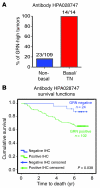
Figure 7. GRN expression correlates with aggressive tumor subtypes and reduced survival of breast cancer patients.
(A) Percentage of tumors in each category (triple-negative [TN]/basal or nonbasal) that scored positively for high GRN staining using antibody HPA028747. (B) Kaplan-Meier analysis of correlation between GRN-positive (green) or GRN-negative (blue) expression and survival.
Figure 8
Figure 8. The systemic instigation model.
Instigating tumors secrete endocrine factors, including but not limited to OPN (9), that mediate the expression of GRN by Sca1+cKitCD45+ hematopoietic cells in the host BM. These activated BMCs are subsequently mobilized into the circulation and are recruited to sites where otherwise indolent responding tumors reside. The GRN-expressing BMCs assume close proximity to tissue fibroblasts within the tumor stroma and induce these fibroblasts to express αSMA as well as genes related to cytokine- and chemokine-mediated inflammation, integrin signaling, and matrix remodeling. This systemic instigation cascade ultimately results in malignant growth of the responding tumors.

"VSports注册入口" Comment in

References

    1. Schoenberg BS. Multiple primary malignant neoplasms. The Connecticut experience, 1935–1964. Recent Results Cancer Res. 1977;58:1–173. - PubMed
    1. Watanabe S, et al. Multiple primary cancers in 5,456 autopsy cases in the National Cancer Center of Japan. J Natl Cancer Inst. 1984;72(5):1021–1027. - PubMed
    1. Schaapveld M, et al. Risk of new primary nonbreast cancers after breast cancer treatment: a Dutch population-based study. J Clin Oncol. 2008;26(8):1239–1246. doi: 10.1200/JCO.2007.11.9081. - DOI - PubMed
    1. Schaapveld M, et al. The impact of adjuvant therapy on contralateral breast cancer risk and the prognostic significance of contralateral breast cancer: a population based study in the Netherlands. Breast Cancer Res Treat. 2008;110(1):189–197. doi: 10.1007/s10549-007-9709-2. - DOI - PMC - PubMed
    1. Carmichael AR, Bendall S, Lockerbie L, Prescott R, Bates T. The long-term outcome of synchronous bilateral breast cancer is worse than metachronous or unilateral tumours. Eur J Surg Oncol. 2002;28(4):388–391. doi: 10.1053/ejso.2002.1266. - DOI - PubMed

Publication types (VSports注册入口)

MeSH terms

Substances