Skip to main page content
U.S. flag

An official website of the United States government

Dot gov

The . gov means it’s official. Federal government websites often end in . gov or VSports app下载. mil. Before sharing sensitive information, make sure you’re on a federal government site. .

Https

The site is secure V体育官网. The https:// ensures that you are connecting to the official website and that any information you provide is encrypted and transmitted securely. .

. 2011 Jan 10;13(1):R2.
doi: 10.1186/bcr2803.

Knockdown of miR-21 in human breast cancer cell lines inhibits proliferation, in vitro migration and in vivo tumor growth

Affiliations

Knockdown of miR-21 in human breast cancer cell lines inhibits proliferation, in vitro migration and in vivo tumor growth (VSports最新版本)

"V体育官网入口" Li Xu Yan et al. Breast Cancer Res. .

V体育ios版 - Abstract

Introduction: MicroRNAs (miRNAs) are a class of small non-coding RNAs (20 to 24 nucleotides) that post-transcriptionally modulate gene expression. A key oncomir in carcinogenesis is miR-21, which is consistently up-regulated in a wide range of cancers. However, few functional studies are available for miR-21, and few targets have been identified VSports手机版. In this study, we explored the role of miR-21 in human breast cancer cells and tissues, and searched for miR-21 targets. .

Methods: We used in vitro and in vivo assays to explore the role of miR-21 in the malignant progression of human breast cancer, using miR-21 knockdown. Using LNA silencing combined to microarray technology and target prediction, we screened for potential targets of miR-21 and validated direct targets by using luciferase reporter assay and Western blot V体育安卓版. Two candidate target genes (EIF4A2 and ANKRD46) were selected for analysis of correlation with clinicopathological characteristics and prognosis using immunohistochemistry on cancer tissue microrrays. .

Results: Anti-miR-21 inhibited growth and migration of MCF-7 and MDA-MB-231 cells in vitro, and tumor growth in nude mice. Knockdown of miR-21 significantly increased the expression of ANKRD46 at both mRNA and protein levels. Luciferase assays using a reporter carrying a putative target site in the 3' untranslated region of ANKRD46 revealed that miR-21 directly targeted ANKRD46. miR-21 and EIF4A2 protein were inversely expressed in breast cancers (rs = -0. 283, P = 0. 005, Spearman's correlation analysis) V体育ios版. .

Conclusions: Knockdown of miR-21 in MCF-7 and MDA-MB-231 cells inhibits in vitro and in vivo growth as well as in vitro migration VSports最新版本. ANKRD46 is newly identified as a direct target of miR-21 in BC. These results suggest that inhibitory strategies against miR-21 using peptide nucleic acids (PNAs)-antimiR-21 may provide potential therapeutic applications in breast cancer treatment. .


Figures
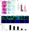
Figure 1
Figure 1
Altered expression of miR-21 in different breast tumor types and breast cell lines. (a) CISH detection using miR-21 LNA detection probe (blue) or scramble-miR as negative control were performed on consecutive 8 μM cryo-sections obtained from BC/FA tissues and corresponding NATs. DNA was counterstained with methyl green (green, × 400 magnification). (b) Total RNA was used to quantify miR-21 expression by relative qRT-PCR, normalizing on U6 RNA levels. The graph shows a log2-scale RQ calculated by normalizing the miR-21 expression values in the tumors on those in the NATs. Data indicate the mean (+SD) of four independent samples. * P < 0.05. (c) FISH detection of miR-21 in five BC cell lines (MCF-7, MDA-MB-231, MDA-MB-453, MDA-MB-435, SK-BR-3) and one non-tumorigenic epithelial cell line (MCF-10A). Positive in situ hybridization signals are visualized in green, while blue depicts DAPI nuclear stain (× 1,000 magnification). (d) qRT-PCR analysis show that MCF-7, MDA-MB-231 and MDA-MB-453 cells express higher levels of miR-21 compared with MCF-10A cells. Data are mean (+SD) of three replicates. ** P < 0.01; BC, breast cancer; FA, fibroadenoma; NATs, normal adjacent tissues.
Figure 2
Figure 2
LNA-antimiR-21 suppressed BC cell growth, proliferation and migration in vitro. (a) miR-21 expression levels (normalized to U6 RNA) were significantly depressed by 98% (P < 0.01) in MCF-7/LNA-antimiR-21 and 77% (P < 0.01) in MDA/LNA-antimiR-21 cells, relative to the LNA-control. The graph shows RQ calculated by normalizing the miR-21 expression values in LNA-antimiR-21 treated cells on those in the LNA-control treated cells. (b) MTT assay showed that MCF-7/antimiR-21 and MDA-MB-231/antimiR-21 cells grew slower than cells transfected with the LNA-control. (c) Representative wells showing total numbers of colonies formed by LNA-antimiR-treated cells standardized against control cells (set to 100%). (d) Representative image of in vitro wound healing assay of MCF-7 and MDA-MB-231 at 0, 24 and 48 h after wound scratch. Data are mean + SD, n = 3. * P < 0.05, ** P < 0.01.
Figure 3
Figure 3
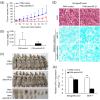
Effect of miR-21-knockdown on MCF-7 cells growth in nude mice: in vivo functional studies. MCF-7 cells (107 cells/tumor), treated with PNA-antimiR-21 or PNA-control (100 nM for 48 h) without transfection reagents, were injected subcutaneously into the left axilla of nude mice. (a) Growth curves for MCF/PNA-antimiR-21 (n = 5) vs. MCF/PNA-control (n = 8) cells in an in vivo proliferation assay. All P-values > 0.05. (b) Tumors were weighed after animals were killed at 17 days post-tumor-cell injection. Decreasing trend for both number and size of tumors from MCF/PNA-antimiR-21 compared with MCF/PNA-control group, although differences were not significant (P = 0.065, Mann-Whitney test). (c) Mice and tumors extracted from MCF/PNA-antimiR-21 and MCF/PNA-control groups. (d) Representative photomicrographs of CISH for miR-21 on xenograft tumor sections obtained from mice bearing MCF/PNA-antimiR-21 or MCF/PNA-control groups (× 400). (e) qRT-PCR analysis of miR-21 expression in MCF-7 cells 48 h post PNA-treatment and in xenograft tumors after mice sacrifice. Data in (a), (b) and (e) indicate the mean + SD. * P < 0.05, ** P < 0.01. PNA, peptide nucleic acid.
Figure 4
Figure 4
mRNA profiling of miR-21-knockdown BC cell lines. MCF-7 and MDA-MB-231 cells were transfected with 50 nM LNA-antimiR-21 or LNA-control. Total RNAs were isolated from MCF-7 cells 48 h post transfection and from MDA-MB-231 cells 36 h post transfection, respectively, and were hybridized to the human genome oligo array V1.0 (CapitalBio, China). (a) The scatter plot shows the LNA-antimiR-21 expression values normalized to LNA-control values, applying a cut-off of 1.3. Each dot represents one probe set. (b) Unsupervised hierarchical cluster analysis of intersection of mRNAs differentially expressed in MCF-7 and MDA-MB-231 cells upon LNA-antimiR-21 treatment. Rows, mRNAs; columns, biological samples. For each mRNA, red is expression higher than average expression across all samples, green is expression lower than average. (c) Validation of microarray results by qRT-PCR, normalizing on GAPDH RNA levels. MCF/A, MCF/LNA-antimiR-21; MCF/C, MCF/LNA-control; MDA/A, MDA/LNA-antimiR-21, MDA/C, MDA/LNA-control.
Figure 5
Figure 5
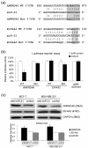
ANKRD46 is a direct target of miR-21 in BC cell lines. (a) Predicted alignment of miR-21 with the target site derived from ANKRD46 and EIF4A2 3' UTR, determined with the software miRBase Targets V5 and miRNAMap, respectively. Note the seed matches at the 5' end of miR-21 (grey boxes) and the mutated nucleotides (underlined). (b) Luciferase assays show that miR-21 directly repress ANKRD46 mRNAs through 3' UTR interactions. Part of the 3' UTRs of wild-type ANKRD46 (478-bp length) and EIF4A2 (194-bp length), or the mutations were cloned into pMIR-REPORT vector (Applied Biosystems), downstream of luciferase. These vectors were then cotransfected with synthetic miR-21 (pre-miR-21) or miR-control in 293T cells, and luciferase activity was quantified. The graph shows the percentage of remaining luciferase activity calculated by normalizing the miR-21 expression values on the miR-control values. (c) Western blot to assay ANKRD46 and EIF4A2 after miR-21 knockdown, with GAPDH as equal loading control, followed by densitometric analysis. Cells were treated and harvested as described in Figure 4b. Data in (b) and (c) are mean + SD (n = 3). * P < 0.05. WT, wild-type; Mut, mutant.
Figure 6
Figure 6
EIF4A2 or ANKRD46 expression and survival in BC patients. (a) Representative TMAs sections stained for ANKRD46 and EIF4A2 from different BC. Examples for staining score 0 (negative), 3, 5 and 7 of marker expression are shown. All images were taken at × 200. (b) Kaplan-Meier survival curve and log-rank test for 99 BC patients showing five-year overall survival. ANKRD46 expression had no significant relationship to patient survival (P = 0.146). High EIF4A2 expression was associated with significantly improved OS (P = 0.044) in women with BC. TMAs, tissue microarrays; OS, overall survival.

References

    1. Parkin DM, Bray F, Ferlay J, Pisani P. Global cancer statistics, 2002. CA Cancer J Clin. 2005;55:74–108. doi: 10.3322/canjclin.55.2.74. - DOI - PubMed
    1. Ambros V. MicroRNA pathways in flies and worms: growth, death, fat, stress, and timing. Cell. 2003;113:673–676. doi: 10.1016/S0092-8674(03)00428-8. - DOI - PubMed
    1. Esquela-Kerscher A, Slack FJ. Oncomirs - microRNAs with a role in cancer. Nat Rev Cancer. 2006;6:259–269. doi: 10.1038/nrc1840. - DOI - PubMed
    1. Chan JA, Krichevsky AM, Kosik KS. MicroRNA-21 is an antiapoptotic factor in human glioblastoma cells. Cancer Res. 2005;65:6029–6033. doi: 10.1158/0008-5472.CAN-05-0137. - "V体育平台登录" DOI - PubMed
    1. Fulci V, Chiaretti S, Goldoni M, Azzalin G, Carucci N, Tavolaro S, Castellano L, Magrelli A, Citarella F, Messina M, Maggio R, Peragine N, Santangelo S, Mauro FR, Landgraf P, Tuschl T, Weir DB, Chien M, Russo JJ, Ju J, Sheridan R, Sander C, Zavolan M, Guarini A, Foa R, Macino G. Quantitative technologies establish a novel microRNA profile of chronic lymphocytic leukemia. Blood. 2007;109:4944–4951. doi: 10.1182/blood-2006-12-062398. - V体育2025版 - DOI - PubMed

MeSH terms