Skip to main page content
U.S. flag

An official website of the United States government

Dot gov

The . gov means it’s official. Federal government websites often end in . gov or . mil. Before sharing sensitive information, make sure you’re on a federal government site. VSports app下载.

Https

The site is secure. The https:// ensures that you are connecting to the official website and that any information you provide is encrypted and transmitted securely V体育官网. .

. 2011 Jan 21;41(2):210-20.
doi: 10.1016/j.molcel.2010.12.005. Epub 2010 Dec 30.

miR-182-mediated downregulation of BRCA1 impacts DNA repair and sensitivity to PARP inhibitors

Affiliations

miR-182-mediated downregulation of BRCA1 impacts DNA repair and sensitivity to PARP inhibitors

Patryk Moskwa et al. Mol Cell. .

Erratum in

  • Mol Cell. 2014 Jan 9;53(1):162-3

Abstract

Expression of BRCA1 is commonly decreased in sporadic breast tumors, and this correlates with poor prognosis of breast cancer patients. Here we show that BRCA1 transcripts are selectively enriched in the Argonaute/miR-182 complex and miR-182 downregulates BRCA1 expression. Antagonizing miR-182 enhances BRCA1 protein levels and protects them from IR-induced cell death, while overexpressing miR-182 reduces BRCA1 protein, impairs homologous recombination-mediated repair, and render cells hypersensitive to IR. The impaired DNA repair phenotype induced by miR-182 overexpression can be fully rescued by overexpressing miR-182-insensitive BRCA1. Consistent with a BRCA1-deficiency phenotype, miR-182-overexpressing breast tumor cells are hypersensitive to inhibitors of poly (ADP-ribose) polymerase 1 (PARP1). Conversely, antagonizing miR-182 enhances BRCA1 levels and induces resistance to PARP1 inhibitor VSports手机版. Finally, a clinical-grade PARP1 inhibitor impacts outgrowth of miR-182-expressing tumors in animal models. Together these results suggest that miR-182-mediated downregulation of BRCA1 impedes DNA repair and may impact breast cancer therapy. .


Figures

Figure 1
Figure 1
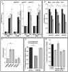
Expression of miRNA cluster-183 is induced during differentiation and suppressed by γ-radiation and BRCA1 is a target of miR-182. A) Expression of miR-183, miR-96 and miR-182 during differentiation of HL60 cells to neutrophils (DMSO, 8 days, p<0.001) and macrophages (TPA, 3 days, p<0.001); K562 cells to megakaryocytes (TPA, 3 days, p<0.002) and to erythrocytes (Hemin, 4 days, p<0.003). B) miR-183, miR-96 and miR-182 are rapidly down-regulated with γ-radiation in proliferating cells. HL60 and K562 cells (data not shown) were exposed to indicate doses of γ-radiation, and RNA isolated 30 min after exposure. There was significant reduction in miR-183 (p<0.002), miR-96 (*p<0.003) and miR-182 (*p<0.002) at 3 Gy. In all the above experiments (panel A and B) the expression of miR-183, miR-96, and miR-182 was analyzed with qRT-PCR and normalized to RNU6B, mean ± SD, n=3-6 independent experiments, are shown. C) Isolation of target transcripts associated with miR-182. Hela cells were co-transfected with expression vectors for HA-tagged AGO1 and miR-182/miR-scr. The immunoprecipitated RNA was analyzed by qRT-PCR using gene-specific primers and normalized to 5S rRNA. FOXO3 and GAPDH served as positive and negative controls, respectively. BRCA1, and not NHEJ1, was significantly (p<0.002) enriched in the pull-down. D, E) miR-182 targets the 3′UTR of BRCA1 mRNA in a luciferase reporter assay. HeLa cells were co-transfected with BRCA1 3′UTR-luciferase reporter, wildtype (WT, stripped) or mutant (M, grey) (D), or with the four predicted miR-182 MREs in the BRCA1 3′UTR (E) and control mimic (miR-scr, black) or miR-182 mimic (stripped and grey) for 48 h before analysis. Firefly luciferase activity of the reporter was normalized to an internal Renilla luciferase control. Mean ± SD of 3 independent experiments are shown. miR-182 significantly (p<0.001) suppresses lucifererase activity from BRCA1-WT reporter but mutation in the miR-182 recognition sites in BRCA1-M rescues this suppression (p<0.001).
Figure 2
Figure 2
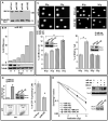
Reduction of BRCA1 protein levels by miR-182 impacts DNA repair. A) miR-182 suppresses BRCA1 expression. HL60 cells transiently transfected with expression vectors for miR-183, miR-96, miR-182 and control (miR-scr), were harvested after 3 days and cell lysates analyzed by immunoblot after normalization for total protein. The indicated non-specific band served as a visual representation for loading control B) Kinetics of miR-182 and BRCA1 protein expression in DMSO-treated HL60 cells. miR-182 expression and BRCA1 protein during DMSO-induced differentiation of HL60 cells. miR-182 was quantified by qRT-PCR normalized to RNU6B. Tubulin served as a loading control for the immunoblot. C) Altering miR-182 levels impacts the amount of unrepaired DSB by comet assay. Proliferating HL60 cells (left panel) were transfected with control or miR-182 mimic and differentiated HL60 cells (right panel) were transfected with control or miR-182 antagomir (ASO). Transfected cells were irradiated, allowed to repair for 18 h and analyzed by single-cell gel electrophoresis (comet assay). BRCA1 protein is compared to tubulin levels in the immunoblots. Representative images are shown in the upper panel and the mean ± SD for each condition below. Residual DNA damage after irradiation is significantly altered in miR-182 mimic (p<0.001) or miR-182 ASO (p<0.001) transfected cells. D) Overexpression of miR-182 impedes homologous recombination-mediated repair of DSBs. U2OS cells carrying the recombination substrate (DR-GFP) were stably transfected with expression vectors for miR-182 or control. I-SceI expression plasmid was transiently transfected and the GFP positive cells analyzed 48 h later by flow cytometry (FACS). miR-182 expression and representative FACS profiles are shown. HR repair was significantly (p<0.002) impaired (lower panel). Mean ± SD of 3 independent experiments is shown. E) BRCA1 mediates the DNA damage sensitivity induced by miR-182. Cells were mock transfected; transfected with miR-182 antagomir (ASO); transfected with miR-182 mimic or BRCA1 cDNA lacking the 3′UTR or both. Cell viability was assayed after indicated doses of γ-radiation by clonogenic cell survival assay. Curves were generated from 3 independent experiments. miR-182 mimic significantly (p<0.003) enhanced sensitivity to IR, whereas miR-182 ASO reduced (p<0.001) IR-sensitivity. Representative immunoblots for D and E are shown.
Figure 3
Figure 3
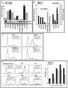
Cell cycle dependent correlation of miR-182 and BRCA1 in breast tumor lines. A, B) miR-182 and BRCA1 protein in breast tumor cell lines. miR-182 quantified by qRT-PCR (normalized to RNU6) and relative to non-tumorigenic breast epithelial cell, HMEC, is shown on the upper panel. Mean ± SD, n=3 independent experiments are shown. A representative immunoblot for BRCA1 (tubulin as control) from the indicated cells are shown in the lower panel. Relative BRCA1 expression was quantified by densitometry using tubulin as control and normalized to expression in HMEC. The estrogen receptor (ER) expression status of the different tumor lines has been indicated. C) Cell cycle profile of the indicated ER-negative cell lines. Asynchronously growing cells were fixed, stained with PI and analyzed by flow cytometry. The cell cycle distribution was assessed using FloJo. Representative images from 3 independent experiments are shown. D) Expression of miR-182 and BRCA1 in synchronized cells. 21NT cells were synchronized using a combination of double thymidine block and nocadozole, and the relative amount of miR-182 and BRCA1 mRNA determined by qRT-PCR (normalized to 5S rRNA). Representative images of the cell cycle profile at indicated times after release are shown on the left panel. Mean ± SD, n=3 independent experiments are shown on the right panel.
Figure 4
Figure 4
miR-182 targets BRCA1 in breast cancer cell lines and impacts function. A) Kinetics of miR-182 and BRCA1 protein expression in TPA-treated MCF7 cells. miR-182 expression and BRCA1 protein levels during TPA-induced differentiation of MCF7 cells. miR-182 was quantified by qRT-PCR normalized to RNU6B. Mean ±SD, n=3 independent experiments, p<0.0091 are shown. Tubulin served as a loading control for the immunoblot. B, C) miR-182 is rapidly down-regulated with IR, and not other DNA damaging agents, in proliferating breast epithelial cells. Proliferating MCF7 cells (B, left panel and C, right panel) and TPA-treated post-mitotic MCF7 cells (B, right panel) were exposed to indicate doses of IR, UV (C, middle panel) and camptothecin (C, left panel) for 1 h. RNA isolated 30 min after exposure. There was significant reduction of miR-182 in MCF7 cells (*p<0.007). The expression of miR-182 was analyzed with qRT-PCR and normalized to RNU6B and in all panels, mean ± SD, n=3-6 independent experiment, are shown. D) Altering miR-182 levels impacts the amount of unrepaired DSB by comet assay in ER negative tumor cells. MDA-MB231 cells (left panel) were transfected with either control mimic (miR-scr) or 182 mimic (miR-182). Conversely, 21NT cells (right panel) were transfected with antagomirs (ASO), either control (AS0-scr) or ASO-182. Transfected cells were irradiated at indicated doses, allowed to repair for 18 h and analyzed by single-cell gel electrophoresis (comet assay). BRCA1 protein is compared to tubulin levels in the immunoblots. Representative images are shown in the upper panel and the mean ±SD, n=3 independent experiments are shown below. Residual DNA damage after irradiation is significantly altered in 182 mimic (miR-182, p<0.001) or ASO-182 (p<0.001) transfected cells.
Figure 5
Figure 5
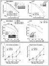
miR-182 mediated regulation of BRCA1 impacts sensitivity to radiation and PARP1 inhibitor in breast cancer cells. A, B) BRCA1 mediates the radiosensitivity, and sensitivity to PARP1 inhibitor, induced by miR-182 in breast cancer cell lines. MDA-MB231 cells (left panels) were transfected with either control mimic or 182 mimic or BRCA1 cDNA lacking the 3′UTR or both. Conversely, 21NT cells (right panels) were transfected with either control ASO or 182 ASO. Cell viability was assayed by clonogenic cell survival assay after indicated doses of γ-radiation (A) or in the presence of PARP1 inhibitor (ANI) at indicated concentrations (B). Curves were generated from 3 independent experiments. miR-182 mimic significantly enhanced sensitivity to IR (p<0.004) and to ANI (p<0.001), whereas miR-182 ASO reduced sensitivity to IR (p<0.001) and ANI (p<0.002). Representative immunoblots for A and B are shown. C) miR-182 impacts PARP inhibitor sensitivity in xenograft mouse models. MDA-MB231 cells stably expressing miR-scr or miR-182 were implanted in each thigh of nude mice (n=5) and olaparib treatment was initiated either 2 days post-implantation (left panel) or tumors outgrown (4-7 weeks) were treated (right panel). Tumor volume was determined in 7 days intervals, and the median fold differences after olaparib treatment represented graphically.

Comment in

References

    1. Atchley DP, Albarracin CT, Lopez A, Valero V, Amos CI, Gonzalez-Angulo AM, Hortobagyi GN, Arun BK. Clinical and pathologic characteristics of patients with BRCA-positive and BRCA-negative breast cancer. J Clin Oncol. 2008;26:4282–4288. - PMC - PubMed
    1. Baldassarre G, Battista S, Belletti B, Thakur S, Pentimalli F, Trapasso F, Fedele M, Pierantoni G, Croce CM, Fusco A. Negative regulation of BRCA1 gene expression by HMGA1 proteins accounts for the reduced BRCA1 protein levels in sporadic breast carcinoma. Mol Cell Biol. 2003;23:2225–2238. - PMC - PubMed
    1. Bandres E, Cubedo E, Agirre X, Malumbres R, Zarate R, Ramirez N, Abajo A, Navarro A, Moreno I, Monzo M, Garcia-Foncillas J. Identification by Real-time PCR of 13 mature microRNAs differentially expressed in colorectal cancer and non-tumoral tissues. Mol Cancer. 2006;5:29. - PMC - PubMed
    1. Bartel DP. MicroRNAs: target recognition and regulatory functions. Cell. 2009;136:215–233. - "VSports手机版" PMC - PubMed
    1. Bhattacharyya A, Ear US, Koller BH, Weichselbaum RR, Bishop DK. The breast cancer susceptibility gene BRCA1 is required for subnuclear assembly of Rad51 and survival following treatment with the DNA cross-linking agent cisplatin. J Biol Chem. 2000;275:23899–23903. - PubMed

MeSH terms

LinkOut - more resources