Skip to main page content
U.S. flag

An official website of the United States government

Dot gov

The . gov means it’s official. Federal government websites often end in . gov or . mil. Before sharing sensitive information, make sure you’re on a federal government site. VSports app下载.

Https

The site is secure V体育官网. The https:// ensures that you are connecting to the official website and that any information you provide is encrypted and transmitted securely. .

. 2010 Dec 21;107(51):22231-6.
doi: 10.1073/pnas.1015245107. Epub 2010 Dec 2.

"VSports app下载" Inositol polyphosphate 4-phosphatase II regulates PI3K/Akt signaling and is lost in human basal-like breast cancers

Affiliations

"VSports注册入口" Inositol polyphosphate 4-phosphatase II regulates PI3K/Akt signaling and is lost in human basal-like breast cancers

Clare G Fedele et al. Proc Natl Acad Sci U S A. .

Abstract

Inositol polyphosphate 4-phosphatase-II (INPP4B) is a regulator of the phosphoinositide 3-kinase (PI3K) signaling pathway and is implicated as a tumor suppressor in epithelial carcinomas. INPP4B loss of heterozygosity (LOH) is detected in some human breast cancers; however, the expression of INPP4B protein in breast cancer subtypes and the normal breast is unknown. We report here that INPP4B is expressed in nonproliferative estrogen receptor (ER)-positive cells in the normal breast, and in ER-positive, but not negative, breast cancer cell lines VSports手机版. INPP4B knockdown in ER-positive breast cancer cells increased Akt activation, cell proliferation, and xenograft tumor growth. Conversely, reconstitution of INPP4B expression in ER-negative, INPP4B-null human breast cancer cells reduced Akt activation and anchorage-independent growth. INPP4B protein expression was frequently lost in primary human breast carcinomas, associated with high clinical grade and tumor size and loss of hormone receptors and was lost most commonly in aggressive basal-like breast carcinomas. INPP4B protein loss was also frequently observed in phosphatase and tensin homolog (PTEN)-null tumors. These studies provide evidence that INPP4B functions as a tumor suppressor by negatively regulating normal and malignant mammary epithelial cell proliferation through regulation of the PI3K/Akt signaling pathway, and that loss of INPP4B protein is a marker of aggressive basal-like breast carcinomas. .


Conflict of interest statement

The authors declare no conflict of interest.

"V体育平台登录" Figures
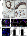
Fig. 1.
Fig. 1.
INPP4B is expressed in nonproliferative ER-positive cells in normal human breast. (A) Normal human breast sections were immunostained using an INPP4B-specific monoclonal antibody (3D5). (Scale bars, 50 μm.) (B) Immunofluorescent images of normal breast sections costained for INPP4B (green), ER (red), and the nuclear marker, ToPro3 iodide (blue) (see also Fig. S2). Coexpression of INPP4B and ER is indicated by arrows. Arrowheads show cells negative for both INPP4B and ER. (Scale bars, 25 μm, 75 μm.) (C) Immunofluorescent costaining of normal human breast lobules for INPP4B (green), Ki-67 (red), and ToPro3 iodide (blue) (Fig. S2). Arrows show INPP4B-positive cells. Outlines and asterisks mark Ki-67–positive nuclei. (Scale bar, 25 μm.) More than 100 Ki-67–positive lobular cells/section were assessed for INPP4B expression, and the mean number of cells that were INPP4B negative (closed bars) or INPP4B positive (open bars) ± SEM from three normal breasts is shown in D. More than 1,000 lobular cells/section were assessed for Ki-67 and INPP4B expression and the mean number of INPP4B-positive lobular cells that were Ki-67 negative (closed bars) or Ki-67 positive (open bars) ± SEM from two normal breasts is shown in E. ***P < 0.002.
Fig. 2.
Fig. 2.
INPP4B protein and mRNA expression in a panel of human breast cancer cells lines. (A) Human breast cancer cell line lysates were immunoblotted for INPP4B, ER, and β-tubulin protein expression, revealing INPP4B protein is expressed in ER-positive, but not ER-negative, cell lines. (B) qRT-PCR analysis of INPP4B mRNA, relative to GAPDH, in ER-positive versus ER-negative human breast cancer cell lines. The graph shows the mean INPP4B mRNA expression ± SEM from two independent experiments.
Fig. 3.
Fig. 3.
Decreased INPP4B expression enhances Akt phosphorylation and tumorigenic potential. (A) INPP4B protein knockdown in two MCF-7-luc-F5 cell populations expressing unique INPP4B-specific shRNAs (INPP4B KD[1] and [2]) was confirmed by immunoblotting. The mean INPP4B protein levels relative to vector control ± SEM from three independent experiments is shown. (B and C) Immunoblot analysis of INPP4B, pSer473-Akt, and total Akt in cells serum-starved for 24 h (B) or stimulated with EGF (C), demonstrating enhanced pSer473-Akt in INPP4B knockdown cells. The mean fold change in pSer473-Akt/total Akt relative to control ± SEM from six (B) or three (C) independent experiments is shown. (D) INPP4B knockdown and control cells were serum starved for 24 and 48 h and assessed for BrdU incorporation as a marker of proliferation. The mean fold increase in BrdU-positive cells relative to control ± SEM from three independent experiments is shown. (E) INPP4B knockdown or control cells were grown in soft agar. The number of colonies/well was determined and the mean fold increase in colony number relative to control ± SEM from two independent experiments performed in triplicate is shown. (F and G) MCF-7-luc-F5 cells expressing control vector or INPP4B-specific shRNAS were injected into the mammary fat pads of Balbc nu/nu mice in the presence of estrogen and tumor growth was analyzed by bioluminescence and caliper measurement. Bioluminescent imaging of xenograft tumors in vivo (F) and mean tumor volumes at 2 and 5.5 wk postinjection (G) indicate INPP4B knockdown cells form larger tumors in vivo. The mean tumor volumes ± SEM of five animals/group is shown. *P < 0.05, **P < 0.01, ***P < 0.005.
Fig. 4.
Fig. 4.
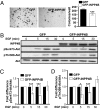
INPP4B protein reconstitution decreases Akt phosphorylation and cell proliferation. (A) INPP4B-null MDA MB 231 cells expressing GFP alone or GFP-tagged INPP4B were suspended in 0.3% agar and colonies allowed to grow for 3 wk. The number of colonies/well was then determined and the mean number of colonies/well ± SEM from two independent experiments performed in triplicate is shown. (B) Immunoblot analysis of INPP4B, pSer473-Akt, pThr308-Akt, and total Akt in GFP or GFP-INPP4B–expressing cells in response to EGF. The mean fold difference in pSer473-Akt (C) and pThr308-Akt (D) normalized to total Akt relative to controls ± SEM from three independent experiments is shown. *P = 0.05, ***P < 0.005.
Fig. 5.
Fig. 5.
INPP4B protein expression in primary human breast cancers. (A) Immunoblot analysis of INPP4B protein expression in a panel of ER-positive and ER-negative primary human breast cancer lysates. ER-negative tumors were predominantly INPP4B-negative (asterisks). (B) Representative IHC staining of INPP4B in primary human breast carcinomas, scored as: 0, no expression; 1, low; 2, moderate; and 3, strong. Inset image in tumor scored 0 for INPP4B shows INPP4B expression in adjacent normal tissue. (Scale bar, 200 μm.) (C) The frequency of INPP4B protein loss of expression in breast cancer subtypes from two independent human datasets [MCCS (black bars) and SVCOC (open bars)] was determined by IHC. Loss of INPP4B was positively correlated with the basal-like subtype and negatively correlated with the luminal A subtype in both datasets. ***P < 0.0001.

References

    1. Ma K, Cheung SM, Marshall AJ, Duronio V. PI(3,4,5)P3 and PI(3,4)P2 levels correlate with PKB/akt phosphorylation at Thr308 and Ser473, respectively; PI(3,4)P2 levels determine PKB activity. Cell Signal. 2008;20:684–694. - PubMed
    1. Franke TF, Kaplan DR, Cantley LC, Toker A. Direct regulation of the Akt proto-oncogene product by phosphatidylinositol-3,4-bisphosphate. Science. 1997;275:665–668. - "V体育官网入口" PubMed
    1. Scheid MP, et al. Phosphatidylinositol (3,4,5)P3 is essential but not sufficient for protein kinase B (PKB) activation; phosphatidylinositol (3,4)P2 is required for PKB phosphorylation at Ser-473: Studies using cells from SH2-containing inositol-5-phosphatase knockout mice. J Biol Chem. 2002;277:9027–9035. - VSports - PubMed
    1. Liu P, Cheng H, Roberts TM, Zhao JJ. Targeting the phosphoinositide 3-kinase pathway in cancer. Nat Rev Drug Discov. 2009;8:627–644. - PMC - PubMed
    1. Norris FA, Majerus PW. Hydrolysis of phosphatidylinositol 3,4-bisphosphate by inositol polyphosphate 4-phosphatase isolated by affinity elution chromatography. J Biol Chem. 1994;269:8716–8720. - PubMed

Publication types

MeSH terms