Skip to main page content
U.S. flag

An official website of the United States government

Dot gov

The . gov means it’s official. Federal government websites often end in VSports app下载. gov or . mil. Before sharing sensitive information, make sure you’re on a federal government site. .

Https

The site is secure. The https:// ensures that you are connecting to the official website and that any information you provide is encrypted and transmitted securely. V体育官网.

. 2011 Mar 24;30(12):1449-59.
doi: 10.1038/onc.2010.526. Epub 2010 Nov 22.

MUC1 enhances invasiveness of pancreatic cancer cells by inducing epithelial to mesenchymal transition

Affiliations

"VSports app下载" MUC1 enhances invasiveness of pancreatic cancer cells by inducing epithelial to mesenchymal transition

L D Roy et al. Oncogene. .

"VSports" Abstract

Increased motility and invasiveness of pancreatic cancer cells are associated with epithelial to mesenchymal transition (EMT). Snai1 and Slug are zinc-finger transcription factors that trigger this process by repressing E-cadherin and enhancing vimentin and N-cadherin protein expression. However, the mechanisms that regulate this activation in pancreatic tumors remain elusive. MUC1, a transmembrane mucin glycoprotein, is associated with the most invasive forms of pancreatic ductal adenocarcinomas (PDA). In this study, we show that over expression of MUC1 in pancreatic cancer cells triggers the molecular process of EMT, which translates to increased invasiveness and metastasis. EMT was significantly reduced when MUC1 was genetically deleted in a mouse model of PDA or when all seven tyrosines in the cytoplasmic tail of MUC1 were mutated to phenylalanine (mutated MUC1 CT). Using proteomics, RT-PCR and western blotting, we revealed a significant increase in vimentin, Slug and Snail expression with repression of E-Cadherin in MUC1-expressing cells compared with cells expressing the mutated MUC1 CT VSports手机版. In the cells that carried the mutated MUC1 CT, MUC1 failed to co-immunoprecipitate with β-catenin and translocate to the nucleus, thereby blocking transcription of the genes associated with EMT and metastasis. Thus, functional tyrosines are critical in stimulating the interactions between MUC1 and β-catenin and their nuclear translocation to initiate the process of EMT. This study signifies the oncogenic role of MUC1 CT and is the first to identify a direct role of the MUC1 in initiating EMT during pancreatic cancer. The data may have implications in future design of MUC1-targeted therapies for pancreatic cancer. .


Conflict of interest statement

Conflict of Interest:

Drs. Mukherjee and Gendler work has been funded by the NIH. There are no other conflicts to declare V体育安卓版. All other authors declare no conflicts of interests.

Figures (VSports app下载)
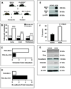
Figure 1
Figure 1. PDA mice lacking Muc1 have significantly lower incidence of secondary metastasis associated with decreased induction of EMT proteins in the tumors
A. Schematic representation of PDA, PDA.MUC1 and PDA.Muc1KO mice. PDA mice were mated to the human MUC1.Tg and mouse Muc1 KO mice to get PDA.MUC1 mice and PDA.MUC1 KO mice respectively. B. Percent mice that developed metastasis in PDA.MUC1 versus PDA.MUC1 KO mice. C and D. Proteomics data on PDA.MUC1 and PDA.Muc1 KO tumor lysates showing E-cadherin repression and Vimentin upregulation. E. Western blot analysis of MUC1 expression for KCM and KCKO cells using MUC1 TR and MUC1 CT monoclonal antibodies. F. Transwell invasion assay showing significantly higher invasion index for KCM cells compared to KCKO cells (*P<0.001). G. Gain of mesenchymal proteins in KCM cells versus KCKO cells. β-actin serves as control for equal protein loading.
Figure 2
Figure 2. Significantly higher invasion and transcription of genes associated with a mesenchymal phenotype coupled with significantly lower transcription of genes associated with an epithelial phenotype in MUC1 over-expressing human pancreatic cancer cells as compared to control cells. Complete reversal in the MUC1.Y0 expressing cells
MUC1 expression by Western blot analysis: A. MUC1 TR and B. MUC1 CT expression of BxPC3 Neo, MUC1, and Y0; C. MUC1 TR and D. MUC1 CT staining of Su86.86 Neo, MUC1, and Y0. >200kDa represents MUC1 TR domain and 30kD represents MUC1 CT domain. β-actin was used as loading control. E - F. In vitro trans-well invasion assay for BxPC3 and Su86.86 cells respectively; Compared to Neo and Y0 cells, MUC1-expressing cells have significantly higher invasion index (* p<0.001). G - H. RT-PCR analysis of BxPC3 Neo, MUC1, and Y0 cells: G. Transcription of genes generally associated with epithelial phenotype. H. Transcription of genes generally associated with mesenchymal phenotype. Genes whose transcription was altered by at least 2-fold or more were considered significant. Average fold change is shown from three separate experiments. All experiments were repeated 3 times with a R-value (Correlation Coefficient) of >0.98. Clones from three independent infections were analyzed with similar results.
Figure 3
Figure 3. Repression of E-Cadherin and induction of Snail, Slug, and Vimentin in MUC1 cells coupled with significantly increased transcription of genes associated with metastasis. Complete reversal in MUC1-Y0 expressing cells
A – D. Western blotting analysis of Slug, Snail, Vimentin (A and C), and E-Cadherin (B and D) expression in BxPC3 and Su86.86 respectively. Neo, MUC1, and Y0 cells are shown. E. Immunofluorescence staining and confocal microscopic image of E-Cadherin levels in BxPC3 Neo, MUC1 and Y0 cells. Images were taken at 400X magnification. The experiments were repeated 3 times with three separate clones with similar results. β-actin was used as loading control. F. RT-PCR analysis of genes generally associated with metastasis and angiogenesis. 2-fold or more difference was considered significant. Average fold change is shown from three separate experiments.
Figure 4
Figure 4. Higher tumor burden with loss of E-Cadherin and gain of mesenchymal and metastatic proteins in BxPC3 MUC1 versus Y0 and Neo tumors
A. In vivo tumor growth in nude mice. Significantly higher tumor burden in BxPC3. MUC1 versus Neo and Y0 tumors (*p < 0.001). B. MUC1 expression by Western blotting using the MUC1 TR and CT antibodies C. Tumor cells cultured from whole blood of tumor-bearing mice. Circulating tumor cells detected in the blood of mice bearing the BxPC3 MUC1 but not BxPC3 Y0 or Neo tumors. D. Expression of E-Cadherin in primary tumors by Western blotting. E. Expression of Slug, Snail, and Vimentin in primary tumors by Western blotting. F. High levels of pro-metastatic and pro-angiogenic proteins detected in the BXPC3 MUC1 tumor lysate. Fold change in levels of various factors in BxPC3 MUC1 tumor lysate compared to Y0 and Neo, G. Expression of VEGF and E-cadherin by IHC. Images were taken at 400X magnification. Similar results with n=3 mice were obtained.
Figure 5
Figure 5. MUC1 interacts with β-catenin and translocates to the nucleus in the MUC1-expressing cells. Complete reversal in the Y0 cells
Protein expression of MUC1 CT and β-catenin in the nuclear extracts of A. BxPC3 MUC1 and Y0; B. KCM and KCKO and C. Su86.86 MUC1 and Y0 cells. Lamin and IKK used as positive and negative control for nuclear extracts. Co-IP of MUC1 CT and β-catenin in both directions from D. BxPC3 MUC1 and Y0; E. KCM and KCKO and F. Su86.86 MUC1 and Y0 cells. Non-specific pull down was not detected using an IgG control antibody. Note: Neo cells were not included in the data as they express minimal levels of endogenous MUC1 and we found little to no β-catenin or MUC1 CT in the nucleus (data not shown)

References

    1. Chang BW, Siccion E, Saif MW. Updates in locally advanced pancreatic cancer. Highlights from the "2010 ASCO Annual Meeting". Chicago, IL, USA. June 4–8, 2010. JOP. 2010;11:313–316. - "V体育官网入口" PubMed
    1. Burris HA, 3rd, Moore MJ, Andersen J, Green MR, Rothenberg ML, Modiano MR, Cripps MC, Portenoy RK, Storniolo AM, Tarassoff P, Nelson R, Dorr FA, Stephens CD, Von Hoff DD. Improvements in survival and clinical benefit with gemcitabine as first-line therapy for patients with advanced pancreas cancer: a randomized trial. J Clin Oncol. 1997;15:2403–2413. - PubMed
    1. Thiery JP. Epithelial-mesenchymal transitions in tumour progression. Nat Rev Cancer. 2002;2:442–454. - PubMed
    1. Nieto MA. The snail superfamily of zinc-finger transcription factors. Nat Rev Mol Cell Biol. 2002;3:155–166. - PubMed
    1. Batlle E, Sancho E, Franci C, Dominguez D, Monfar M, Baulida J, Garcia De Herreros A. The transcription factor snail is a repressor of E-cadherin gene expression in epithelial tumour cells. Nat Cell Biol. 2000;2:84–89. - PubMed

Publication types

MeSH terms