<code lang="qMgMiFw"></code> Skip to main page content
U.S. flag

An official website of the United States government

Dot gov

The . gov means it’s official. Federal government websites often end in . gov or . mil VSports app下载. Before sharing sensitive information, make sure you’re on a federal government site. .

Https

The site is secure. The https:// ensures that you are connecting to the official website and that any information you provide is encrypted and transmitted securely. V体育官网.

Comparative Study
. 2010 Dec 14;49(49):10421-39.
doi: 10.1021/bi1012518. Epub 2010 Nov 15.

Characterization of nicotinamidases: steady state kinetic parameters, classwide inhibition by nicotinaldehydes, and catalytic mechanism

Affiliations
Comparative Study

Characterization of nicotinamidases: steady state kinetic parameters, classwide inhibition by nicotinaldehydes, and catalytic mechanism

"VSports手机版" Jarrod B French et al. Biochemistry. .

Abstract

Nicotinamidases are metabolic enzymes that hydrolyze nicotinamide to nicotinic acid. These enzymes are widely distributed across biology, with examples found encoded in the genomes of Mycobacteria, Archaea, Eubacteria, Protozoa, yeast, and invertebrates, but there are none found in mammals. Although recent structural work has improved our understanding of these enzymes, their catalytic mechanism is still not well understood. Recent data show that nicotinamidases are required for the growth and virulence of several pathogenic microbes. The enzymes of Saccharomyces cerevisiae, Drosophila melanogaster, and Caenorhabditis elegans regulate life span in their respective organisms, consistent with proposed roles in the regulation of NAD(+) metabolism and organismal aging. In this work, the steady state kinetic parameters of nicotinamidase enzymes from C. elegans, Sa. cerevisiae, Streptococcus pneumoniae (a pathogen responsible for human pneumonia), Borrelia burgdorferi (the pathogen that causes Lyme disease), and Plasmodium falciparum (responsible for most human malaria) are reported. Nicotinamidases are generally efficient catalysts with steady state k(cat) values typically exceeding 1 s(-1). The K(m) values for nicotinamide are low and in the range of 2 -110 μM VSports手机版. Nicotinaldehyde was determined to be a potent competitive inhibitor of these enzymes, binding in the low micromolar to low nanomolar range for all nicotinamidases tested. A variety of nicotinaldehyde derivatives were synthesized and evaluated as inhibitors in kinetic assays. Inhibitions are consistent with reaction of the universally conserved catalytic Cys on each enzyme with the aldehyde carbonyl carbon to form a thiohemiacetal complex that is stabilized by a conserved oxyanion hole. The S. pneumoniae nicotinamidase can catalyze exchange of (18)O into the carboxy oxygens of nicotinic acid with H(2)(18)O. The collected data, along with kinetic analysis of several mutants, allowed us to propose a catalytic mechanism that explains nicotinamidase and nicotinic acid (18)O exchange chemistry for the S. pneumoniae enzyme involving key catalytic residues, a catalytic transition metal ion, and the intermediacy of a thioester intermediate. .


Figures (VSports app下载)
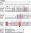
Figure 1
Figure 1
Multiple sequence alignment of the Pyrococcus horikoshii (PDB entry 1IM5), Streptococcus pneumoniae, Escherichia Coli, Saccharomyces cerevisiae, and Borrelia burgdorferi nicotinamidases. The alignment was performed with ClustalW (56) and edited with ESPript (57). Identical residues are highlighted, and the secondary structure elements of the solved P. horikoshii structure (33) are shown above the alignment. Vertical arrows point to the proposed catalytic triad proposed by Du et al. that is composed of an aspartate, cysteine, and lysine residue. The proposed metal-binding residues are shown with an M under the appropriate residue
Figure 2
Figure 2
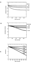
(A) The GDH assay was performed with varying concentrations of ammonium acetate. (B) The GDH-coupled assay was performed with varying concentrations of nicotinamide. Reactions containing 1 mM α-ketoglutarate, 250 µM of NADPH, 1.5 units GDH per 100 µL of reaction volume in 100 mM phosphate buffer pH 7.3 were initiated with the addition of PfNic to a final concentration of 320 nM. The reactions were measured by monitoring the absorbance at 340 nm over time. (C) SpNic activity scales nicely with varying concentrations of enzyme. Reactions containing 1 mM α-ketoglutarate, 250 µM of NADPH, 1.5 units GDH per 100 µL of reaction volume in 100 mM phosphate buffer pH 7.3 were initiated with the addition of SpNic to the amounts indicated in the figure. The reactions were measured by monitoring the absorbance at 340 nm over time. The slopes for 0, 36, 72 and 108 pmol of SpNic are −2.23 × 10⁻⁵, −2.39 × 10⁻⁴, −4.79 × 10⁻⁴ and −7.30 × 10⁻⁴, respectively.
Figure 3
Figure 3
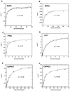
The steady state saturation curves of nicotinamidases from S. cerevisiae (A), B. burgdorferi (B), P. falciparum (C), and S. pneumoniae (D), C. elegans PNC1 (E) and C. elegans PNC2 (F). A and D were derived from progress curve analysis (see experimental) of the nicotinamidase reaction run in the presence of 100 µM nicotinamide, B and C are from HPLC analysis of reactions run in varying nicotinamide concentrations, E and F are from the GDH coupled fluorescence assay with various concentrations of nicotinamide. In all cases, the data was fit to the Michaelis-Menten equation using KaleidaGraph.
Figure 4
Figure 4
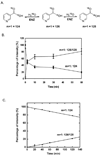
Mass spectra demonstrating time dependent 18O exchange into nicotinic acid (NA) catalysed by SpNic. Data is plotted as percent intensity versus time, where the total intensities of peaks in mass spectra at m/z = 124, 126, and 128 are summed to reach 100 percent. 124 is the mass of unlabeled nicotinic acid as a twice protonated cation (M+). 126 is the molecular mass of nicotinic acid incorporating a single 18O label, and 128 is the mass of nicotinic acid incorporating two 18O atoms. Mass intensities of single and double-labeled nicotinic acid are summed. The data in panel B shows that the mass spectrum of nicotinic acid treated with SpNic in the presence of 18O becomes rapidly depleted of intensity for the peak corresponding to m/z = 124, whereas there is a corresponding enrichment in m/z = 126 and m/z = 128 peak intensities, demonstrating 18O incorporation into nicotinic acid. Control does not exchange. A. Schematic exchange reaction catalyzed by SpNic. B. Time course of 18O exchange catalyzed by SPNic. The reaction was carried out in 1 mM NA, 18O-H2O, 6 µM SPNic at 37 °C for the indicated time. C. Determination of the initial rate of 18O exchange. The reaction was carried out in 1 mM NA, 18O-H2O, 100 nM SpNic at 37 °C for the indicated time. The initial rate was determined to be 0.34 s−1 after correction for the mole fraction of 18O present in the reaction (Equation 4, Materials and Methods).
Figure 5
Figure 5
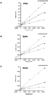
The Lineweaver-Burke plots for nicotinamidases from P. falciparum (A), S. pneumoniae (B) and B. burgdorferi (C) in the presence of nicotinaldehyde determined by fluorescence plate reader assay. Curves were obtained in the presence of three different concentrations of nicotinaldehyde, demonstrating the competitive nature of the inhibitor with substrate. The units in the y-axis are expressed as s/A.U. (A.U.: arbitrary units). A control showing that nicotinaldehyde does not interfere with the GDH enzyme activity is provided in the Supporting Information.
Figure 6
Figure 6
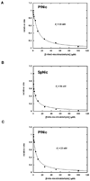
Curves showing the inhibition of the nicotinamidase enzymes by 5-O-methylnicotinaldehyde (A, [PfNic] = 14 nM) and 5-methylnicotinaldehyde (B, [SpNic] = 12 nM C, [PfNic] = 14 nM). The reactions were run in the presence of 1 mM nicotinamide and various concentrations of the inhibitor in 1 mM α-ketoglutarate, 250 µM NADH and 1.5 units of GDH per 100 µL of reaction volume in 100 mM phosphate buffer, pH 7.3. After initiation by addition of the enzyme, the reactions were monitored by fluorescence and initial rates of reaction were calculated. The curves show observed rate with respect to inhibitor concentration. Data points are fit to Morrisons equation (39) and the Ki calculated from the Kiapp value of Morrison’s equation as described in the Materials and Methods. Controls showing that nicotinaldehydes do not interfere with the GDH enzyme activity is provided in the Supporting Information.
Scheme 1
Scheme 1
Nicotinamidase hydrolyzes nicotinamide to give nicotinic acid.
Scheme 2
Scheme 2
Nicotinamidase catalyzes the conversion of pyrazinamide to pyrazinoic acid.
Scheme 3
Scheme 3
The nicotinamidase assay developed couples the release of ammonia to the consumption of NAD(P)H via glutamate dehydrogenase. The synthesis of glutamate by GDH occurs in the presence of ammonia and α-ketoglutarate and is dependent upon the stoichiometric oxidation of NAD(P)H. The conversion of NAD(P)H to NAD(P)+ can be followed by monitoring the decrease in absorbance at 340 nm or the decrease in fluorescence (excitation 360 nm, emission 490 nm).
Scheme 4
Scheme 4
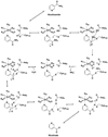
Mechanism for nicotinaldehyde inhibition of nicotinamidase enzymes. Numbering is from the enzyme from SpNic. Top figure represents reaction coordinate diagram, where plateaus levels are meant to indicate relative energy.
Scheme 5
Scheme 5
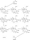
Mechanism for nicotinic acid reversal to the nicotinoyl thioester complex. The mechanism explains the ability of SpNic to catalyse 18O exchange into nicotinic acid. A key feature of the proposed mechanism is the formation of a deprotonation of Lys103 upon thioester formation from nicotinic acid, which then serves as a base catalyst to activate water in the 18O exchange reaction. The numbering system used is for the SpNic enzyme.
Scheme 6
Scheme 6
Proposed mechanism of nicotinamidases. The ammonia generated in the first half of the reaction coordinate generates deprotonated Lys residue prior to departure from the active site. The intermediacy of this Lys is envisioned to act as a base catalyst that activates a water to complete the second half of the catalytic reaction. The numbering system used is for the SpNic enzyme.
Scheme 7
Scheme 7
Alternative aqua-Zn+2 mechanism of nicotinamidases. The ammonia generated in the first half of the reaction coordinate generates a Zn-hydroxide by deprotonation prior to departure from the active site. The intermediacy of a Zn-hydroxide is envisioned to act as a base catalyst that activates a solvent derived nucleophilic water in the second half of the catalytic reaction. The numbering system used is for the SpNic enzyme.

References

    1. Gerdes SY, Scholle MD, D'Souza M, Bernal A, Baev MV, Farrell M, Kurnasov OV, Daugherty MD, Mseeh F, Polanuyer BM, Campbell JW, Anantha S, Shatalin KY, Chowdhury SA, Fonstein MY, Osterman AL. From genetic footprinting to antimicrobial drug targets: examples in cofactor biosynthetic pathways. J Bacteriol. 2002;184:4555–4572. - "V体育官网入口" PMC - PubMed
    1. Boshoff HI, Mizrahi V. Purification, gene cloning, targeted knockout, overexpression, and biochemical characterization of the major pyrazinamidase from Mycobacterium smegmatis. J Bacteriol. 1998;180:5809–5814. - PMC - PubMed
    1. Zhang H, Deng JY, Bi LJ, Zhou YF, Zhang ZP, Zhang CG, Zhang Y, Zhang XE. Characterization of Mycobacterium tuberculosis nicotinamidase/pyrazinamidase. Febs J. 2008;275:753–762. - VSports app下载 - PubMed
    1. Scorpio A, Zhang Y. Mutations in pncA, a gene encoding pyrazinamidase/nicotinamidase, cause resistance to the antituberculous drug pyrazinamide in tubercle bacillus. Nat Med. 1996;2:662–667. - PubMed
    1. Ghislain M, Talla E, Francois JM. Identification and functional analysis of the Saccharomyces cerevisiae nicotinamidase gene, PNC1. Yeast. 2002;19:215–224. - PubMed

Publication types

MeSH terms

"VSports最新版本" Substances

LinkOut - more resources