Skip to main page content (V体育官网)
U.S. flag

An official website of the United States government

Dot gov

The . gov means it’s official. Federal government websites often end in . gov or VSports app下载. mil. Before sharing sensitive information, make sure you’re on a federal government site. .

Https

The site is secure. The https:// ensures that you are connecting to the official website and that any information you provide is encrypted and transmitted securely V体育官网. .

Review
. 2010 Jun;15(2):169-90.
doi: 10.1007/s10911-010-9181-1. Epub 2010 May 15.

The pathophysiology of epithelial-mesenchymal transition induced by transforming growth factor-beta in normal and malignant mammary epithelial cells

Affiliations
Review

The pathophysiology of epithelial-mesenchymal transition induced by transforming growth factor-beta in normal and malignant mammary epithelial cells (V体育官网)

Molly A Taylor et al. J Mammary Gland Biol Neoplasia. 2010 Jun.

Abstract

Epithelial-mesenchymal transition (EMT) is an essential process that drives polarized, immotile mammary epithelial cells (MECs) to acquire apolar, highly migratory fibroblastoid-like features. EMT is an indispensable process that is associated with normal tissue development and organogenesis, as well as with tissue remodeling and wound healing. In stark contrast, inappropriate reactivation of EMT readily contributes to the development of a variety of human pathologies, particularly those associated with tissue fibrosis and cancer cell invasion and metastasis, including that by breast cancer cells. Although metastasis is unequivocally the most lethal aspect of breast cancer and the most prominent feature associated with disease recurrence, the molecular mechanisms whereby EMT mediates the initiation and resolution of breast cancer metastasis remains poorly understood VSports手机版. Transforming growth factor-beta (TGF-beta) is a multifunctional cytokine that is intimately involved in regulating numerous physiological processes, including cellular differentiation, homeostasis, and EMT. In addition, TGF-beta also functions as a powerful tumor suppressor in MECs, whose neoplastic development ultimately converts TGF-beta into an oncogenic cytokine in aggressive late-stage mammary tumors. Recent findings have implicated the process of EMT in mediating the functional conversion of TGF-beta during breast cancer progression, suggesting that the chemotherapeutic targeting of EMT induced by TGF-beta may offer new inroads in ameliorating metastatic disease in breast cancer patients. Here we review the molecular, cellular, and microenvironmental factors that contribute to the pathophysiological activities of TGF-beta during its regulation of EMT in normal and malignant MECs. .


Figures

Figure 1
Figure 1
Schematic depicting the canonical and noncanonical TGF-β signaling systems coupled to EMT in MECs. Transmembrane signaling by TGF-β ensues through its binding and activation of the Ser/Thr protein kinase receptors, TβR-I and TβR-II. Indeed, TGF-β binding to either TβR-III or TβR-II enables the recruitment and transphosphorylation of TβR-I, resulting its activation and subsequent phosphorylation the receptor-activated Smads, Smad2 and Smad3. Once activated, Smad2/3 form heterocomplexes with common the Smad, Smad4, which collectively translocate en masse to the nucleus to regulate the expression of TGF-β-responsive genes in concert with an ever expanding list of transcriptional coactivators and repressors. This branch of the bifurcated TGF-β signaling system represents the “canonical” or “Smad2/3-dependent” TGF-β pathway (left). Alternatively, TGF-β also activates a variety of “noncanonical” or “Smad2/3-independent” effectors, including Par6, NF-κB, ILK, FAK, Src, a variety of small GTPases, members of the MAP kinase family (e.g., ERK1/2, JNK, and p38 MAPK), and the PI3K:AKT:mTOR signaling axis (right). Activation of members of the Snail, ZEB, or bHLH family of transcription factors promote EMT by transcriptionally repressing the production of E-cadherin transcripts (bottom). Imbalances between canonical and noncanonical TGF-β signaling pathways have recently been identified and established as casual aberrancies underlying the initiation of oncogenic TGF-β signaling and its initiation of EMT in normal and malignant MECs. See text for details.
Figure 2
Figure 2
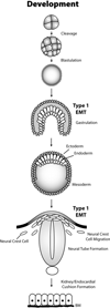
Type 1 EMT in development and embryogenesis. Following fertilization, the zygote undergoes several rounds of cleavage to form a dense spheroid structure, which then undergoes blastulation to from a hollowed blastula. During gastrulation, TGF-β provides inductive EMT signals that elicits the formation of an invaginating structure that generates the three multipotent germ layers, namely the mesoderm, endoderm, and ectoderm. The mesoderm gives rise to muscle, skeleton, and connective tissue, while the endoderm gives rise to internal organs, such as the liver, colon, and stomach. The outer ectodermal layer generates the epidermis and ocular lens, as well as produces the mammary gland. The ectoderm also gives rise to the primordial nervous system via neurulation and neural crest formation, a process that dependent upon EMT induced by TGF-β. Finally, TGF-β stimulation of EMT during organogenesis is essential for the faithful development of the kidney, and of the endocardial cushion and subsequent atrioventricular valve formation. The role of TGF-β in regulating type 1 EMT during mammary gland branching and invasion is discussed in the text.
Figure 3
Figure 3
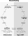
Type 2 EMT in wound healing and tissue regeneration. Normal epithelia that arose from type 1 EMT during development experience a noxious event or injury that triggers endothelial and epithelial cells to produce factors that mediate coagulation and clot formation. These events, coupled with enhanced MMP production facilitate the recruitment of immune cells and platelets to denuded wounds, leading to the production of provisional ECM and activation of angiogenesis. TGF-β enhances the healing process by inducing EMT in myofibroblasts, which drives their differentiation, activation, and migration into denuded wounds to facilitate wound restitution, closure, and re-epithelialization. The role of TGF-β in regulating type 2 EMT during mammary gland fibrosis is discussed in the text.
Figure 4
Figure 4
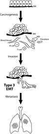
Type 3 EMT in cancer metastasis. Normal epithelia that arose from type I EMT during development experience a carcinogenic event that ultimately results in their oncogenic transformation and tumor formation. The development and progression of mammary tumors is accompanied by their acquisition of dysplastic and abnormal morphologies, and by their evolution in chronically inflamed tumor microenvironments, which further enhances their acquisition of EMT and fibrotic phenotypes. Through its ability to stimulate oncogenic EMT, TGF-β enables transitioned mammary carcinoma cells to invade the underlying basement membrane (BM) and escape the confines of the primary tumor. Once liberated, metastatic MECs undergo intravasation and traverse the blood stream prior to taking up residence at distant locales, an event that ultimately leads to disease recurrence and poor clinical outcomes in breast cancer patients. See text for specific details on the molecular mechanisms whereby TGF-β promotes type 3 EMT and metastasis in breast cancer cells.

References

    1. Wendt MK, Allington TM, Schiemann WP. Mechanisms of the epithelial-mesenchymal transition by TGF-β. Future Oncol. 2009;5(8):1145–1168. - PMC - PubMed
    1. Kalluri R, Weinberg RA. The basics of epithelial-mesenchymal transition. J Clin Invest. 2009;119(6):1420–1428. - "V体育安卓版" PMC - PubMed
    1. Thiery JP, Acloque H, Huang RY, Nieto MA. Epithelial-mesenchymal transitions in development and disease. Cell. 2009;139(5):871–890. - PubMed
    1. Yang J, Weinberg RA. Epithelial-mesenchymal transition: at the crossroads of development and tumor metastasis. Dev Cell. 2008;14(6):818–829. - PubMed
    1. Hugo H, Ackland ML, Blick T, Lawrence MG, Clements JA, Williams ED, et al. Epithelial--mesenchymal and mesenchymal--epithelial transitions in carcinoma progression. J Cell Physiol. 2007;213(2):374–383. - PubMed

Publication types

MeSH terms

Substances (V体育2025版)