"V体育平台登录" Skip to main page content
U.S. flag

An official website of the United States government

Dot gov

The . gov means it’s official. Federal government websites often end in . gov or . mil. Before sharing sensitive information, make sure you’re on a federal government site VSports app下载. .

Https

The site is secure. The https:// ensures that you are connecting to the official website and that any information you provide is encrypted and transmitted securely. V体育官网.

. 2010 May 3;5(5):e10443.
doi: 10.1371/journal.pone.0010443.

Jnk2 effects on tumor development, genetic instability and replicative stress in an oncogene-driven mouse mammary tumor model

Affiliations

Jnk2 effects on tumor development, genetic instability and replicative stress in an oncogene-driven mouse mammary tumor model

"VSports" Peila Chen et al. PLoS One. .

Abstract (V体育官网)

Oncogenes induce cell proliferation leading to replicative stress, DNA damage and genomic instability. A wide variety of cellular stresses activate c-Jun N-terminal kinase (JNK) proteins, but few studies have directly addressed the roles of JNK isoforms in tumor development. Herein, we show that jnk2 knockout mice expressing the Polyoma Middle T Antigen transgene developed mammary tumors earlier and experienced higher tumor multiplicity compared to jnk2 wildtype mice. Lack of jnk2 expression was associated with higher tumor aneuploidy and reduced DNA damage response, as marked by fewer pH2AX and 53BP1 nuclear foci. Comparative genomic hybridization further confirmed increased genomic instability in PyV MT/jnk2-/- tumors. In vitro, PyV MT/jnk2-/- cells underwent replicative stress and cell death as evidenced by lower BrdU incorporation, and sustained chromatin licensing and DNA replication factor 1 (CDT1) and p21(Waf1) protein expression, and phosphorylation of Chk1 after serum stimulation, but this response was not associated with phosphorylation of p53 Ser15 VSports手机版. Adenoviral overexpression of CDT1 led to similar differences between jnk2 wildtype and knockout cells. In normal mammary cells undergoing UV induced single stranded DNA breaks, JNK2 localized to RPA (Replication Protein A) coated strands indicating that JNK2 responds early to single stranded DNA damage and is critical for subsequent recruitment of DNA repair proteins. Together, these data support that JNK2 prevents replicative stress by coordinating cell cycle progression and DNA damage repair mechanisms. .


Conflict of interest statement

Competing Interests: The authors have declared that no competing interests exist.

Figures

Figure 1
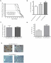
Figure 1. Systemic jnk2 deletion enhances tumor development.
PyV MT/jnk2+/+ (n = 12), PyV MT/jnk2+/− (n = 16), and PyV MT/jnk2−/− (n = 19) mice were palpated for mammary tumors thrice weekly. Once palpated, tumor growth was recorded thrice weekly. A). Kaplan Meier graph showing age of first tumor palpation (median age was day 55 for PyV MT/jnk2−/− vs. day 70 for PyV MT/jnk2+/+, p = .11); B). Total number of tumors palpated per mouse at the time of harvest was higher in PyV MT/jnk2−/− mice compared to PyV MT/jnk2+/+ mice, p = 0.0192); C). Paraffin embedded, non-target tumor sections were probed with cleaved caspase 3 primary antibody and detected using FITC labeled secondary antibody. Nuclei were stained with propidium iodide. The total number of cells staining positive for cleaved caspase 3 were scored and divided by the total number of nuclei (n = 5 tumors in each group); D). Paraffin embedded tissue sections were probed with Ki-67 primary antibody and detected using DAB. Cells staining positive for Ki-67 were counted and divided by the total number of nuclei (Hematoxylin) per field. Five fields per tumor were counted (n = 5 per genotype, p = 0.0159); E). Paraffin embedded tissue sections were probed with p-c-Jun (Ser63) primary antibody and detected using DAB. Hematoxylin was used as a nuclear stain.
Figure 2
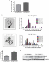
Figure 2. Target tumors obtained from PyV MT/jnk2−/− had increased aneuploidy.
A). Tumors were finely minced and digested as described in the Methods section. Cells were trypsinized at passage 2 to 3 and assessed for DNA content using PI staining. PyV MT/jnk2−/− tumors contained more cells with DNA content ≥4N (PyV MT/jnk2+/+ (n = 5), PyV MT/jnk2−/− (n = 10), p = 0.0485); B). Primary cells were treated with colcemid and harvested to assess number of chromosomes per metaphase. The number and frequency of abnormal (aneuploid) chromosome numbers are higher in PyV MT/jnk2−/− compared to PyV MT/jnk2+/+ (p = 0.0043). Each colored bar represents a single target tumor obtained for an individual mouse. The number to the right of the mouse number denotes the number of metaphases counted for each target tumor. The graph illustrates the frequency of a particular chromosome number for each target tumor; C). Tumor lysates were subjected to SDS PAGE and western blotting using p53 primary antibody and detected using chemiluminescence. GAPDH primary antibody was used to assess similar loading amongst samples.
Figure 3
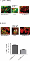
Figure 3. pH2AX and 53BP1 staining in PyV MT tumors.
A). Paraffin embedded PyV MT tumors were stained with pH2AX antibody (green). Nuclei were counter stained with propidium iodide (PI, red); Inset shows high magnification of a cell from a PyV MT/jnk2+/+ tumor with arrows pointing at foci. B). Tumor sections were processed as in A) except incubated with 53BP1 primary antibody. Images were captured and pseudo-colored. 53BP1 antibody (red) and DAPI nuclear stain (green). Inset shows high magnification of a cell from the PyV MT/jnk2+/+ with arrows pointing at foci. C). Cells with ≥4 nuclear 53BP1 foci were counted as positive cells in each field; a minimum of 5 fields were counted/mouse. Each point represents a tumor average (p = 0.0286).
Figure 4
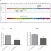
Figure 4. Chromosomal and gene expression changes occur in PyV MT/jnk2−/− tumors.
A). aCGH analysis of PyV MT/jnk2−/− tumors show deletions in segments containing lig1, anacp5 and amplifications in dmp-1, amongst others, compared to PyV MT/jnk2+/+ controls; B). Expression differences between PyV MT/jnk2+/+ and PyV MT/jnk2−/− tumors were compared for lig1 and anapc5 using qPCR amplification and standard curves (n = 5 per genotype).
Figure 5
Figure 5. Serum treatment of G1 arrested cells induces cell death in PyV MT/jnk2−/− cells.
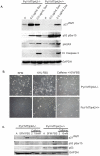
A). PyVMT/jnk2+/+ and PyVMT/jnk2−/− cells were serum starved for 24 hours and then treated with 10% FBS containing medium. Serum stimulated cells were pulsed with BrdU two hours prior to harvesting and then stained with BrdU primary antibody followed by BrdU detection using flow cytometry. BrdU positivity data are presented as percent positive cells in total cell population; B). PyVMT/jnk2+/+ and PyVMT/jnk2−/− cells were serum starved for 24 hours and then treated with 10% FBS containing medium. After 24 hours of serum starvation, cells were either cultured in fresh SFM or medium containing 10% FBS and harvested 24 hours later. Cells were evaluated for Annexin positivity using flow cytometry. Data are expressed as percent positive cells of the total population; C). Cells were serum starved as above and then harvested at indicated time points after 10% FBS stimulation to assess expression of various cell cycle associated proteins using western blot analysis with primary antibodies directed towards the indicated proteins. GAPDH was used to compare even sample loading.
Figure 6
Figure 6. PyV MT/jnk2−/− cellular response is specific to replication and reversed by ATM/ATR inhibitor caffeine.
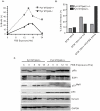
A). PyVMT/jnk2+/+ and PyVMT/jnk2−/− cells were treated with doxorubicin at the indicated concentrations for 18 hours and then lysed. Expression of p21Waf1, p-p53 (Ser15), pH2AX (Ser139), and cleaved caspase 3 were evaluated using western blot analysis. GAPDH was used to compare even loading amongst samples; B). Cells were treated as described in A). except caffeine 2 mM was added as indicated; C). Cells were treated as described in A). except caffeine 2 and 10 mM were added as indicated. Expression of p21Waf1, p-p53 (Ser 15), and p53 were evaluated using western blot analysis. GAPDH was used to compare even loading amongst samples.
Figure 7
Figure 7. PyV MT/jnk2−/− cells experience replicative stress and increased p21Waf1 expression.
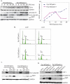
A). Cells were serum starved and then harvested at different time points after 10% FBS stimulation to evaluate CDT1, p21Waf1, p-Chk1 (Ser345), and p-p53 (Ser 15) expression by western blot analysis using primary antibodies directed towards the indicated proteins. CDT1 expression at each time point was normalized to GAPDH and graphed for PyVMT/jnk2+/+ and PyVMT/jnk2−/− cell lines; B). PyVMT/jnk2+/+ and PyVMT/jnk2−/− cell lines were infected with either adenoviral-GFP or adenoviral-CDT1. Forty-eight hours later, cells were stained using PI with RNase, and then evaluated for cell cycle distribution using flow cytometry; C). Cells were infected with either adenoviral-GFP or adenoviral-CDT1 and harvested 24 hours later. Alternatively, cells were treated with hydroxyurea (HU 5 mM) for 24 hours and then harvested. Western blot analysis was used to measure pChk1 (Ser 345) and p21Waf1 expression. GAPDH was used to compare sample loading; D). Cells were infected with either adenoviral-GFP or adenoviral-CDT1 during 24 hours of serum starvation then stimulated with 10% FBS and harvested 24 hours later. pChk1 (Ser 345), p-p53 (Ser15), and p21Waf1 expression was evaluated using western blot analysis. GAPDH was used to compare sample loading.
Figure 8
Figure 8. JNK2 is integral in sensing replicative stress and localizing at RPA coated lesions.
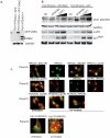
A). PyVMT/jnk2−/− cells were infected with JNK2α retrovirus and selected using puromycin. GFP-JNK2 expression was measured using JNK2 primary antibody and PyVMT/jnk2+/+ lysates as positive a control; B). PyVMT/jnk2−/− and PyVMT/jnk2−/−GFP-JNK2a expressing cells were infected with increasing doses of GFP-CDT1. Cells were processed as described in C). Cell lysates were analyzed for pChk1 (Ser 345), p53 (Ser 15) and p21Waf1. GAPDH was used to compare even sample loading. C). MCF10A cells were plated in chamber slides, untreated or treated with UV (10 J/m2), and fixed 2 hrs later. Cells were incubated with RPA, DNA Ligase 1 (Lig1), PCNA, or JNK2 primary antibodies, as indicated, followed by incubation with FITC or Texas Red secondary antibodies, (G) Green, (R) Red. Panel D includes images acquired using confocal microscopy. Co-localization was evaluated using color overlay.

References

    1. Courtois-Cox S, Jones SL, Cichowski K. Many roads lead to oncogene-induced senescence. Oncogene. 2008;27:2801–2809. - PubMed
    1. Srinivas S, Schonthal A, Eckhart W. Polyomavirus Middle-Sized Tumor Antigen modulates c-Jun phosphorylation and transcriptional activity. Proc Natl Acad Sci U S A. 2007;91:10064–10068. - PMC - PubMed
    1. Rodrigues GA, Morag P, Schlessinger J. Activation of the JNK pathway is essential for transformation by the Met oncogene. EMBO J. 1997;16:2634–2645. - V体育官网入口 - PMC - PubMed
    1. MacLaren A, Black EJ, Clark W, Gillespie DA. c-Jun-deficient cells undergo premature senescence as a result of spontaneous DNA damage accumulation. Molecular & Cellular Biology. 2004;24:9006–9018. - PMC - PubMed
    1. Lin EY, Jones JG, Li P, Zhu L, Whitney KD, et al. Progression to malignancy in the polyoma middle T oncoprotein mouse breast cancer model provides a reliable model for human diseases. American Jorunal of Pathology. 2003;163:2113–2126. - PMC - PubMed

"VSports" Publication types

MeSH terms (VSports最新版本)