Skip to main page content
U.S. flag

An official website of the United States government

Dot gov

The . gov means it’s official. Federal government websites often end in . gov or . mil. Before sharing sensitive information, make sure you’re on a federal government site VSports app下载. .

Https

The site is secure. The https:// ensures that you are connecting to the official website and that any information you provide is encrypted and transmitted securely V体育官网. .

. 2010 Jun;176(6):2948-57.
doi: 10.2353/ajpath.2010.090963. Epub 2010 Apr 29.

Macrophage inhibitory cytokine-1 regulates melanoma vascular development

Affiliations

Macrophage inhibitory cytokine-1 regulates melanoma vascular development

Sung Jin Huh et al. Am J Pathol. 2010 Jun.

Abstract

Expression of macrophage inhibitory cytokine-1 (MIC-1), a member of the transforming growth factor-beta family, normally increases during inflammation or organ injury VSports手机版. MIC-1 is also expressed at higher levels in melanomas; however, its role in tumorigenesis is unknown. This report identifies a novel function for MIC-1 in cancer. MIC-1 was overexpressed in approximately 67% of advanced melanomas, accompanied by fivefold to six-fold higher levels of secreted protein in serum of melanoma patients compared with normal individuals. Constitutively active mutant (V600E)B-Raf in melanoma regulated downstream MIC-1 expression. Indeed, small-interfering RNA-mediated targeting of MIC-1 or (V600E)B-Raf reduced expression and secretion by three-fold to fivefold. This decrease in MIC-1 levels reduced melanoma tumorigenesis by approximately threefold, but did not alter cultured cell growth, suggesting a unique function other than growth control. Instead, inhibition of MIC-1 was found to mechanistically retard melanoma tumor vascular development, subsequently affecting tumor cell proliferation and apoptosis. This role in melanoma angiogenesis was confirmed by comparing MIC-1 and vascular endothelial growth factor (VEGF) function in chick chorioallantoic membrane and matrigel plug assays. Similar to VEGF in melanomas, MIC-1 stimulated directional vessel development, acting as a potent angiogenic factor. Thus, MIC-1 is secreted from melanoma cells together with VEGF to promote vascular development mediated by (V600E)B-Raf signaling. .


Figures

Figure 1
Figure 1
MIC-1 is overexpressed in and secreted from melanomas. A: MIC-1 protein was expressed at higher levels in melanoma patient tumors compared with normal human melanocytes. α-Enolase served as a control for equal protein loading. B: MIC-1 concentrations are elevated in the blood serum of melanoma patients compared with normal individuals. MIC-1 concentrations in blood serum of melanoma patients were measured by ELISA. Values are presented as mean ± SEM. C: MIC-1 is overexpressed in the majority of melanoma cell lines. Expression of MIC-1 was examined in melanoma cell lines established from primary tumors at the radial (RGP; WM35 and WM3211), vertical (VGP; WM115, WM98.1, and WM278), and metastatic (MM; UACC 903) stages of melanoma progression. Only WM3211 did not express MIC-1 at levels higher than that observed in melanocytes. Normal skin melanocytes, fibroblasts, and keratinocytes did not contain detectable levels of MIC-1 protein by Western blotting. α-Enolase was used as a control for equal protein loading. D: MIC-1 was secreted from metastatic melanoma cell lines but not at significant levels from RGP or VGP cell lines. MIC-1 concentrations in media were measured by ELISA. Values are presented as mean ± SEM.
Figure 2
Figure 2
Targeting mutant V600EB-Raf reduced levels of MIC-1 protein expression in and secretion from melanoma cells. SiRNA-mediated inhibition of mutant V600EB-Raf significantly reduced MIC-1 protein expression in UACC 903 (A) and A375M (B) melanoma cells compared with untransfected controls or cells nucleofected with buffer or scrambled siRNA. Reduction of mutant V600EB-Raf protein expression decreased MIC-1 levels similar to that observed after inhibition of MIC-1 expression by using siRNA. α-Enolase was used as a control for equal protein loading. Inhibition of mutant V600EB-Raf reduced MIC-1 secretion from UACC 903 (C) and A375M (D) melanoma cells. Media alone were used as a negative control. Values are presented as mean ± SEM. ***P < 0.001 compared with controls.
Figure 3
Figure 3
Decreasing MIC-1 expression does not alter the growth of cultured melanoma cells. A: SiRNA-mediated knockdown of MIC-1 protein expression did not affect the growth of cultured melanoma cells. UACC 903 or A375M cell viability was measured by using an MTS assay after siRNA-mediated knockdown of MIC-1. Values are presented as mean ± SEM. V600EB-Raf protein knockdown was used as a positive control for inhibition of cell growth. B: Ectopic expression of HA-tagged-V600EB-Raf led to a 50% increase in secretion of MIC-1 from melanoma cells. HA-tagged-V600EB-Raf (5.0, 7.5, and 10 μg of construct) was ectopically expressed in C8161 melanoma cells lacking V600EB-Raf. Western blotting shows expression of HA-tagged protein. α-Enolase was used as a control for equal protein loading. ELISA showed a 50% increase in MIC-1 secretion from cells expressing HA-tagged-V600EB-Raf compared with controls. Values are presented as mean ± SEM. *P < 0.05 compared with controls.
Figure 4
Figure 4
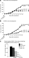
SiRNA-mediated inhibition of MIC-1 retarded the tumorigenic potential of melanoma cells. Targeting MIC-1 by using siRNA inhibits melanoma tumor development. SiRNA targeting MIC-1, siScrambled, or buffer controls were nucleofected into UACC 903 (A) or A375M (B) cell lines, and 36 hours later, cells were subcutaneously injected into nude mice. Tumor size was measured on alternate days up to day 19.5. Values are presented as mean ± SEM. C: siRNA-mediated inhibition reduced MIC-1 levels in mouse blood serum by 50%. At day 19.5 after nucleofection of siRNA targeting MIC-1, levels secreted from A375M tumors were measured from five separate mice by ELISA. Mice lacking xenografted tumors were used as negative control. Values are presented as mean ± SEM. *P < 0.05; ***P < 0.001 compared with controls.
Figure 5
Figure 5
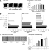
Targeting MIC-1 reduced the angiogenic potential of melanoma cells to decrease melanoma tumor development. A: SiRNA-mediated inhibition of MIC-1 in A375M or UACC 903 tumors reduced protein expression by 50% in time and size matched tumors. B: siRNA-mediated inhibition of MIC-1 in A375M or UACC 903 tumors reduced serum levels of the protein in mice bearing day 13 time and sized matched tumors by 50% (*P < 0.05; **P < 0.01 compared with controls). C: SiRNA-mediated inhibition of MIC-1 reduced vascular development to decrease tumor development. Comparison of vascular development, proliferation, and cellular apoptosis rates in size and time matched UACC 903 tumors differing in MIC-1 protein expression. Change in vascular development was the first statistically significant difference (**P < 0.01) observed in size and time matched tumors in which MIC-1 was targeted by using siRNA. Reduced cell proliferation (*P < 0.05; **P < 0.01) and increased apoptosis (**P < 0.01) were consistently observed starting at day 11. Columns represent means of 12 fields analyzed from four tumors per group; bars represent ± SEM.
Figure 6
Figure 6
MIC-1 and VEGF promote melanoma vascular development. A: MIC-1 increased blood vessels formation in the direction of filter paper soaked with MIC-1. Quantification of newly formed blood vessels showed MIC-1 promoted angiogenesis in the CAM assay. A minimum of 20 filter papers were photographed (original magnification, ×40) and quantified to determine blood vessel number. PBS served as negative control. Values are presented as mean ± SEM. *P < 0.05; **P < 0.01; ***P < 0.001. B: Targeting MIC-1 and/or VEGF suppressed blood vessel formation compared with controls in the chick chorioallantoic membrane assay. SiRNA was used to decrease secreted levels of MIC-1 and/or VEGF and conditioned media used on a CAM assay. Values are presented as mean ± SEM. C: MIC-1 and VEGF stimulation alone and in combination increased formation of blood vessels in the matrigel plug assay. Matrigel was mixed with 2.5 or 5.0 ng/ml of MIC-1 and/or 200 pg/ml VEGF and injected subcutaneously into mice. After 10 days, matrigel plugs were removed, fixed, and photographed (original magnification, ×40). Blood vessels formed in the gels were quantified. Values are presented as mean ± SEM. *P < 0.05 compared with controls.

References

    1. Massague J. TGFbeta in cancer. Cell. 2008;134:215–230. - PMC - PubMed
    1. Leivonen SK, Kahari VM. Transforming growth factor-beta signaling in cancer invasion and metastasis. Int J Cancer. 2007;121:2119–2124. - PubMed
    1. Bootcov MR, Bauskin AR, Valenzuela SM, Moore AG, Bansal M, He XY, Zhang HP, Donnellan M, Mahler S, Pryor K, Walsh BJ, Nicholson RC, Fairlie WD, Por SB, Robbins JM, Breit SN. MIC-1, a novel macrophage inhibitory cytokine, is a divergent member of the TGF-beta superfamily. Proc Natl Acad Sci USA. 1997;94:11514–11519. - PMC - PubMed
    1. Fairlie WD, Moore AG, Bauskin AR, Russell PK, Zhang HP, Breit SN. MIC-1 is a novel TGF-beta superfamily cytokine associated with macrophage activation. J Leukoc Biol. 1999;65:2–5. - PubMed
    1. Hsiao EC, Koniaris LG, Zimmers-Koniaris T, Sebald SM, Huynh TV, Lee SJ. Characterization of growth-differentiation factor 15, a transforming growth factor beta superfamily member induced following liver injury. Mol Cell Biol. 2000;20:3742–3751. - PMC - PubMed

Publication types

MeSH terms

Substances