Skip to main page content
U.S. flag

An official website of the United States government

Dot gov

The . gov means it’s official. Federal government websites often end in . gov or . mil VSports app下载. Before sharing sensitive information, make sure you’re on a federal government site. .

Https

The site is secure V体育官网. The https:// ensures that you are connecting to the official website and that any information you provide is encrypted and transmitted securely. .

. 2010 Mar 8;188(5):629-38.
doi: 10.1083/jcb.200905059. Epub 2010 Mar 1.

Regulators of cyclin-dependent kinases are crucial for maintaining genome integrity in S phase

Affiliations

Regulators of cyclin-dependent kinases are crucial for maintaining genome integrity in S phase

Halfdan Beck et al. J Cell Biol. .

Abstract

Maintenance of genome integrity is of critical importance to cells VSports手机版. To identify key regulators of genomic integrity, we screened a human cell line with a kinome small interfering RNA library. WEE1, a major regulator of mitotic entry, and CHK1 were among the genes identified. Both kinases are important negative regulators of CDK1 and -2. Strikingly, WEE1 depletion rapidly induced DNA damage in S phase in newly replicated DNA, which was accompanied by a marked increase in single-stranded DNA. This DNA damage is dependent on CDK1 and -2 as well as the replication proteins MCM2 and CDT1 but not CDC25A. Conversely, DNA damage after CHK1 inhibition is highly dependent on CDC25A. Furthermore, the inferior proliferation of CHK1-depleted cells is improved substantially by codepletion of CDC25A. We conclude that the mitotic kinase WEE1 and CHK1 jointly maintain balanced cellular control of Cdk activity during normal DNA replication, which is crucial to prevent the generation of harmful DNA lesions during replication. .


Figures (V体育官网)
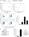
Figure 1.
Figure 1.
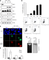
High-throughput siRNA screen identifies WEE1 as a regulator of genomic integrity. (A) Schematic overview of the robot-automated high-throughput siRNA screen. U2OS cells were reverse transfected, and after incubation for 72 h, cells were stained with anti–γ-H2AX antibody and Hoechst. (B) Individual siRNAs were ranked according to the percentage of cells that were γ-H2AX positive (left), and genes were ranked according to their RSA-derived p-value (right). The positions of WEE1 and CHK1 are indicated. (C) U2OS cells were transfected with three different siRNAs targeting WEE1 and harvested for flow cytometric analysis after 48 h. The bar plot is the mean of the percentage of γ-H2AX–positive cells of three experiments. (D) Immunoblot of cells treated as in C. (E) TIG-3-tert cells were transfected with WEE1 siRNA for 48 h and analyzed by flow cytometry and immunoblotting. (C and E) Error bars indicate SD from three experiments. (D and E) Molecular mass is indicated in kilodaltons. PCNA, proliferating cell nuclear antigen.
Figure 2.
Figure 2.
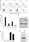
WEE1 depletion leads to DNA damage in newly replicated DNA. (A) U2OS cells were transfected with WEE1 siRNA and processed for immunoblotting. Molecular mass is indicated in kilodaltons. PCNA, proliferating cell nuclear antigen. (B) Flow cytometry analysis for γ-H2AX and propidium iodide (PI) of cells 12, 24, 36, and 48 h after transfection with WEE1 siRNA. The boxed areas indicate γ-H2AX–positive cells.The bar plot is the mean of the percentage of γ-H2AX–positive cells of three experiments. (C) Immunofluorescence confocal microscopy of cells 12 and 18 h after transfection with WEE1 siRNA. To detect ssDNA, cells were cultured in medium with 10 µM BrdU throughout the experiment; DNA was not denatured before staining against BrdU, γ-H2AX, and DAPI. (D) For the detection of replication-associated DSBs, cells were labeled with methyl-[14C]thymidine (14C-TdR) for 30 min. Control and WEE1 samples were labeled 12 h after siRNA transfection. Cells were harvested 24 h after transfection, and DNA fragments were separated by PFGE. IR, ionizing radiation. (E) U2OS cells were depleted for the DNA replication proteins MCM2 and CDT1 for 48 h. WEE1 was depleted the last 20 h. Cells were processed for flow cytometry analysis with staining of γ-H2AX and DNA (PI). (B and E) Error bars indicate SD from three experiments. Bar, 10 µm.
Figure 3.
Figure 3.
DNA damage accumulating after WEE1 depletion is dependent on CDK1 and -2 but not CDC25A. (A) U2OS cells were transfected with WEE1 siRNA with and without 100 nM CDK1 or -2 siRNA for 36 h or with 25 µM of the Cdk inhibitor roscovitine (Rosco) added 12 h after transfection. Cells were processed for flow cytometry analysis with staining of γ-H2AX and DNA (PI). (B) Mean of three experiments treated as in A. (C) Immunoblot of cells treated as in A. (D) U2OS cells were transfected with a WEE1 and CDC25A siRNA pool (50 nM of each siRNA), harvested after 24 h and stained for γ-H2AX and with PI and analyzed by flow cytometry. (B and D) Error bars indicate SD from three experiments. (E) Immunoblot of cells treated as in D. (C and E) Molecular mass is indicated in kilodaltons.
Figure 4.
Figure 4.
Endogenously arising DNA damage after CHK1 depletion is dependent on CDC25A. (A) U2OS cells were transfected with CHK1 siRNA and a CDC25A siRNA pool (50 nM of each siRNA). After 48 h, cells were fixed, stained for γ-H2AX and with PI, and analyzed by flow cytometry. The bar plot is the mean of the percentage of γ-H2AX–positive cells of three experiments. (B) Cells were treated as in A and prepared for immunoblotting. Molecular mass is indicated in kilodaltons. (C) TIG-3-tert cells were transfected with the indicated siRNA and, 24 h later, trypsinized and seeded. Cells were counted at the indicated time. Data are the mean of three experiments. (D) TIG-3-tert cells were transfected with CHK1 siRNA and a CDC25A siRNA pool and, after 24 h, 2 mM HU was added. Confocal microscopy was performed on cells immunostained with an antibody against RAD51 and DAPI for DNA. (E) Quantification of D. 100 cells were counted in triplicates. (A, C, and E) Error bars indicate SD from three experiments. Bars, 5 µm.

VSports - Comment in

References (V体育ios版)

    1. Bader G.D., Betel D., Hogue C.W. 2003. BIND: the Biomolecular Interaction Network Database. Nucleic Acids Res. 31:248–250 10.1093/nar/gkg056 - DOI - PMC - PubMed
    1. Bartek J., Lukas J. 2007. DNA damage checkpoints: from initiation to recovery or adaptation. Curr. Opin. Cell Biol. 19:238–245 10.1016/j.ceb.2007.02.009 - DOI - PubMed
    1. Bartkova J., Rezaei N., Liontos M., Karakaidos P., Kletsas D., Issaeva N., Vassiliou L.V., Kolettas E., Niforou K., Zoumpourlis V.C., et al. 2006. Oncogene-induced senescence is part of the tumorigenesis barrier imposed by DNA damage checkpoints. Nature. 444:633–637 10.1038/nature05268 - DOI - PubMed
    1. Ben-Yehoyada M., Gautier J., Dupré A. 2007. The DNA damage response during an unperturbed S-phase. DNA Repair (Amst.). 6:914–922 10.1016/j.dnarep.2007.02.005 - DOI - PubMed
    1. Chen Y., Poon R.Y. 2008. The multiple checkpoint functions of CHK1 and CHK2 in maintenance of genome stability. Front. Biosci. 13:5016–5029 - PubMed

V体育ios版 - Publication types

MeSH terms