Skip to main page content
U.S. flag

An official website of the United States government

Dot gov

The VSports app下载. gov means it’s official. Federal government websites often end in . gov or . mil. Before sharing sensitive information, make sure you’re on a federal government site. .

Https

The site is secure. The https:// ensures that you are connecting to the official website and that any information you provide is encrypted and transmitted securely. V体育官网.

. 2010 Apr 30;285(18):13561-8.
doi: 10.1074/jbc.M109.075945. Epub 2010 Feb 20.

Myristoylated Naked2 antagonizes Wnt-beta-catenin activity by degrading Dishevelled-1 at the plasma membrane

Affiliations

V体育官网入口 - Myristoylated Naked2 antagonizes Wnt-beta-catenin activity by degrading Dishevelled-1 at the plasma membrane

Tianhui Hu et al. J Biol Chem. .

"V体育ios版" Abstract

In Drosophila, naked cuticle is an inducible antagonist of the Wnt-beta-catenin pathway, likely acting at the level of Dishevelled (Dsh/Dvl), an essential component of this pathway. The mechanism by which naked cuticle and its two vertebrate orthologs, Naked1 (NKD1) and Naked2 (NKD2), inhibit Dvl function is unknown. NKD2 is myristoylated, a co-translational modification that leads to its plasma membrane localization. In contrast, myristoylation-deficient G2A NKD2 is cytoplasmic. Herein we show that the ability of Nkd2/NKD2 to antagonize Wnt-beta-catenin activity during zebrafish embryonic development and in mammalian HEK293 cells is myristoylation-dependent. NKD2 and Dvl-1 interact and co-localize at the lateral membrane of polarized epithelial cells. In reciprocal overexpression and siRNA knockdown experiments, NKD2 and Dvl-1 destabilize each other via enhanced polyubiquitylation; this effect is also dependent upon Naked2 myristoylation. Cell fractionation and ubiquitylation assays indicate that endogenous NKD2 interacts with a slower migrating, ubiquitylated form of Dvl-1 in plasma membrane fractions VSports手机版. These results provide a mechanism by which NKD2 antagonizes Wnt signaling: myristoylated NKD2 interacts with Dvl-1 at the plasma membrane, and this interaction leads to their mutual ubiquitin-mediated proteasomal degradation. .


Figures

FIGURE 1.
FIGURE 1.
NKD2 antagonizes zebrafish embryonic development in a myristoylation-dependent manner. A, two days post-fertilization, zebrafish embryos were injected at the one-cell stage with wnt8 (25 pg) mRNA, wnt8 plus Nkd2a (800 pg) mRNAs or wnt8 plus G2A Nkd2a (800 pg) mRNA and then assayed 2 days post-fertilization for the presence or absence of eyes. B, quantification of results from panel A. Results are expressed as the overall mean ± S.D. of five separate experiments. ***, p < 0.0001 for the ability of Nkd2a to rescue the Wnt8 eyeless phenotype. C, levels of Nkd2 by Western blotting from the four experimental conditions in A using a rabbit anti-Nkd2a serum that is described under “Experimental Procedures.” The 50-kDa band represents full-length Nkd2; the nature of the smaller, ∼30-kDa band is uncertain but may represent an N-terminal proteolytic fragment. α-Actin was used as a loading control.
FIGURE 2.
FIGURE 2.
NKD2, but not G2A NKD2, antagonizes Wnt-β-catenin signaling in HEK293 cells and this effect is Dvl-1 dependent. A, HEK293 cells were co-transfected with 100 ng of superTOPflash (or FOPflash as control), 10 ng of TK-Renilla, 50 ng of Wnt3a, and 50 ng of NKD2 or G2A NKD2 cDNAs or Dvl-1 siRNA. The assays were carried out as described under “Experimental Procedures.” The results shown are from a representative experiment that was performed in triplicate and repeated at least 10 times. Results are expressed as the mean ± S.D. B, efficient knockdown of Dvl-1 protein by Western blotting of HEK293 cells treated with Dvl-1 siRNA.
FIGURE 3.
FIGURE 3.
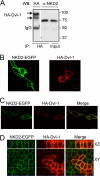
NKD2 and Dvl-1 interact and co-localize at the plasma membrane. A, HEK293 cells were co-transfected transiently with NKD2-EGFP and HA-Dvl-1 cDNAs. Cells were immunoprecipitated with an anti-HA antibody and immunoblotted using both anti-HA and anti-NKD2 (R44) antibodies. B, NKD2-EGFP and HA-Dvl-1 were expressed in MDCK cells individually. NKD2-EGFP localized to cytoplasmic vesicles and the plasma membrane, whereas HA-Dvl-1 decorated scattered puncta in the cytoplasm. C, HA-Dvl-1 co-localized with NKD2-EGFP at the plasma membrane and in some cytoplasmic puncta in MDCK cells transiently co-expressing these cDNAs. D, polarized MDCK cells stably expressing NKD2-EGFP were transfected transiently with HA-Dvl-1. Cells were immunostained and visualized as described under “Experimental Procedures.”
FIGURE 4.
FIGURE 4.
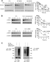
NKD2 and Dvl-1 accelerate each other's degradation via polyubiquitylation. A, MDCK cells were transfected transiently with HA-Dvl-1 and wild-type NKD2-EGFP or G2A NKD2-EGFP. Cycloheximide (CHX, 10 μg/ml) was added for the times indicated to block protein synthesis. Dvl-1 stability was monitored by Western blotting. α-Tubulin served as a loading control. Densitometry was performed, and the intensity of Dvl-1 band was determined after normalization to α-tubulin. B, MDCK cells stably expressing wild-type NKD2-EGFP were transfected transiently with HA-Dvl-1 and control vector (upper panel) or Dvl-1 siRNA and control vector (lower panel). The stability of NKD2 was determined over time following the administration of CHX (10 μg/ml). Levels of NKD2 and α-tubulin were monitored by Western blot analysis. Densitometry was performed, and the intensity of the NKD2 band was determined after normalization to α-tubulin. In A and B, protein levels were normalized to α-tubulin and graphed with time = 0 arbitrarily set to 100%. C, increased ubiquitylation of Dvl-1 (left) or NKD2 (right) in HEK293 cells transiently expressing HA-Ub, FLAG-Dvl-1, or wild-type NKD2. HEK293 cells were treated with MG132 (50 μm) for 4 h before the ubiquitylation assay.
FIGURE 5.
FIGURE 5.
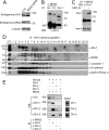
NKD2 specifically recognizes an ubiquitylated form of Dvl-1 at the plasma membrane. A, siRNA knockdown of NKD2 in Caco-2-TGFα cells results in an increased level of Dvl-1 by Western blotting. B, Dvl-1 and NKD2 interaction. Caco-2-TGFα cells were immunoprecipitated with anti-NKD2 antibodies (N-terminal (N-t) R44 or C-terminal (C-t) VU308 NKD2 antibodies) or anti-Dvl-1 antibody and Western blotted with anti-Dvl-1 antibody. C, Caco-2 cells were treated with or without MG132 (50 μm) for 4 h and then lysed, immunoprecipitated with anti-NKD2 antibody, and immunoblotted using anti-Dvl-1 antibody. D, Caco-2-TGFα cells were fractionated over a discontinuous 10–40% iodixanol gradient as described under “Experimental Procedures.” Fractions were blotted with anti-Dvl-1, anti-NKD2, anti-caveolin-1, E-cadherin, and anti-Na+/K+ ATPase α1 antibodies. Slow migrating form of Dvl-1 is found (indicated by arrow) predominantly in early plasma membrane fractions. The additional forms of Dvl-1 detected are similar to those observed from immunoprecipitation and Western blotting of Dvl-1 in B. E, cDNAs of Dvl-1, -2, and -3 and NKD2-EGFP were transiently transfected with a His-tagged ubiquitin cDNA into HEK293T cells. Immunoprecipitations were performed with indicated antibodies. Immunoprecipitates were resolved in the PAGE gel and immunoblotted with anti-ubiquitin and anti-Dvl-1 antibodies. A single slower migrating Dvl-1 band was detected with anti-ubiquitin antibody. Equivalent levels of Dvl-1, -2, and -3 protein were observed by Western blotting in cells transiently transfected with the respective cDNAs (marked as Input in the panel on the right).

References (VSports最新版本)

    1. Cadigan K. M., Nusse R. (1997) Genes Dev. 11, 3286–3305 - PubMed
    1. Wallingford J. B., Habas R. (2005) Development 132, 4421–4436 - "VSports手机版" PubMed
    1. Gan X. Q., Wang J. Y., Xi Y., Wu Z. L., Li Y. P., Li L. (2008) J. Cell Biol. 180, 1087–1100 - PMC - PubMed
    1. Glinka A., Wu W., Delius H., Monaghan A. P., Blumenstock C., Niehrs C. (1998) Nature 391, 357–362 - PubMed
    1. Takemaru K., Yamaguchi S., Lee Y. S., Zhang Y., Carthew R. W., Moon R. T. (2003) Nature 422, 905–909 - V体育安卓版 - PubMed

Publication types

MeSH terms