"V体育安卓版" Skip to main page content
U.S. flag

An official website of the United States government

Dot gov

The . gov means it’s official. Federal government websites often end in . gov or . mil. Before sharing sensitive information, make sure you’re on a federal government site. VSports app下载.

Https

The site is secure. The https:// ensures that you are connecting to the official website and that any information you provide is encrypted and transmitted securely. V体育官网.

. 2010 May 7;285(19):14701-10.
doi: 10.1074/jbc.M110.104711. Epub 2010 Feb 18.

The YTH domain is a novel RNA binding domain

Affiliations

The YTH domain is a novel RNA binding domain

Zhaiyi Zhang et al. J Biol Chem. .

Abstract (V体育平台登录)

The YTH (YT521-B homology) domain was identified by sequence comparison and is found in 174 different proteins expressed in eukaryotes. It is characterized by 14 invariant residues within an alpha-helix/beta-sheet structure. Here we show that the YTH domain is a novel RNA binding domain that binds to a short, degenerated, single-stranded RNA sequence motif. The presence of the binding motif in alternative exons is necessary for YT521-B to directly influence splice site selection in vivo. Array analyses demonstrate that YT521-B predominantly regulates vertebrate-specific exons. An NMR titration experiment identified the binding surface for single-stranded RNA on the YTH domain. Structural analyses indicate that the YTH domain is related to the pseudouridine synthase and archaeosine transglycosylase (PUA) domain. Our data show that the YTH domain conveys RNA binding ability to a new class of proteins that are found in all eukaryotic organisms VSports手机版. .


"VSports" Figures
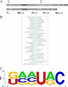
FIGURE 1.
FIGURE 1.
RNA motif binding to YT521-B determined by in vitro SELEX. A, schematic representation of the protein structure of YT521-B. The various protein domains are indicated. E-rich: glutamic-acid rich domain; YTH: YT homology domain (6); P-rich: proline-rich domain; ER-rich: glutamic-acid/arginine-rich domain. 1–4 are the four nuclear localization sites. P1 and P2 are the protein regions used to generate peptide antisera against YT521-B, P1: RSARSVILIFSVRESGKFQCG; P2: KDGELNVLDDILTEVPEQDDECG. B, representative SELEX sequences. The common sequence motif is highlighted in green. C, weight matrix describing the degenerate sequence element present in all SELEX clones.
FIGURE 2.
FIGURE 2.
Interaction of YT521-B with RNA. A, gel mobility shift analysis of YT521-B using RNA probes containing the YTH binding motif. 20 μm of recombinant, baculovirus-generated YT521-B and YT521-B lacking the YTH domain (YTHdel) were incubated with 15 μm YTH binding motif RNA probe. HeLa NE, HeLa nuclear extract serving as a positive control. Probe RNA: GCAUGC. B, gel mobility shift analysis of YT521-B using RNA probes lacking the YTH binding motif. 20 μm of recombinant, baculovirus-generated YT521-B and YT521-B lacking the YTH domain (YTHdel) were incubated with 15 μm control RNA probe (ctlRNA) which is a CUUACU sequence lacking YTH binding motif. C, gel mobility shift analysis of YT521-B using DNA probes. 20 μm of recombinant, baculovirus-generated YT521-B and YT521-B lacking the YTH domain (YTHdel) were incubated with 15 μm single-stranded DNA probe. Probe DNA: GCATGC.
FIGURE 3.
FIGURE 3.
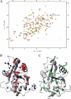
NMR analysis of the YTH domain. A, overlay of the 15N-HSQC spectra of the free domain (red) and in a 2:1 RNA-protein complex (green). A change in position or disappearance of a peak indicates a change in the chemical environment of the respective atoms due to direct binding of the corresponding residue or structural rearrangements upon binding. The identity of selected residues is indicated (sc: side chain). B, backbone or side-chain amides of residues, which either disappeared (red spheres) or showed a large chemical shift changes (blue spheres), were mapped on the NMR structure of the YTH domain solved by the Riken Structural Genomics and Proteomics Initiative (PDB code: 2YUD). These include besides the backbone amides also side-chain atoms from two asparagines and one tryptophan. C, residues proposed to be involved in RNA binding mapped on the structure of the YTH domain.
FIGURE 4.
FIGURE 4.
Influence of YT521-B binding motifs on alternative splicing in vivo. A, structure of the SXN minigene (28). The construct consists of globin exon 1 and 2 (shaded) that flank a central artificial exon. The RNA sequence is introduced in the central exon, indicated by “motif.” Arrows indicate the localization of the primers used for RT-PCR. At least three independent experiments were evaluated using the Student's t test. B, cotransfection assay using a minigene MG-YT1 with the seq1 sequence that contains one YT-521-B binding motif, which is underlined (seq1: AGAGTCCAGTCTGTCAGTCA) sequence. A minigene containing this sequence was cotransfected with an increasing amount of YT521-B or YT521-B (YTHdel). The resulting RNA was analyzed by RT-PCR using the primers indicated in A, p < 0.001. C, cotransfection assay using a minigene MG-YT2 with the seq2 sequence that contains two YTH binding motifs (seq2: GATGCATGCAATGGATGCGG), p < 0.01. D, cotransfection assay using a minigene containing a control sequence seq3 lacking the YTH binding motif (seq3: GGCGATAATGTGTAAATGCC). E, Western blot detecting expression of transfected YT521-B and YT521-B (YTHdel) in cell lysates of the transfection assays. An antibody against EGFP was used for detection. The relative levels of endogenous and transfected YT521-B are shown in Fig. 6C, lane 1.
FIGURE 5.
FIGURE 5.
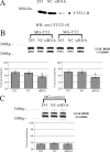
Effect of decreasing YT521-B concentration by siRNA on splice site selection. A, Western blot of the cellular lysates after siRNA treatment. NC, siRNA against pBluescript; siRNA, removal of YT521-B by siRNA. The detection was with an antiserum against YT521-B (7). B, representative ethidium bromide-stained polyacrylamide gels showing the effect of YT521-B reduction by siRNA on the MG-YT1, and MG-YT2 minigenes. 1 μg of the minigenes was cotransfected with the indicated siRNAs in HEK293 cells. C, control, untreated HEK293 cells; NC, siRNA against pBluescript; siRNA, removal of YT521-B by siRNA. The statistical evaluation of three independent experiments is shown underneath representative ethidium-bromide stained gels. C, representative ethidium bromide-stained polyacrylamide gels showing the effect of YT521-B reduction by siRNA on the control minigene (MG-control). The minigene is SXN-based, but the alternative exon does not contain a YTH binding signature. The statistical evaluation is shown underneath the gel.
FIGURE 6.
FIGURE 6.
Mutational analysis of the YTH domain. A, sequence of the YTH domain found in YT521-B (6). The region corresponds to amino acids 358–495. Starting from amino acid 360, every 10th amino acid is indicated with a dash. The phylogenetically conserved residues are indicated by bold letters. The regions predicted to form α helices or β strands are indicated. α-helix: double line, β-sheet: single line. Residues important for influencing splice site selection are circled. Residues that contact RNA are shaded. B, analysis of the MG-YT2 minigene in cotransfection experiments with YTH domain mutations that are indicated by the amino acids changed. p values are 0.0092 (W380D), 0.0038 (F412D), and 0.007 (G414I). C, expression analysis of YTH domain mutations by Western blot, using an antiserum against YT521-B (7). D, analysis of three YTH-domain mutations most severely affecting the YTH domain. 1–3 μg of expression plasmids for each construct were transfected with the MG-YT2 reporter minigene and analyzed by RT-PCR. Parental vector is always included to give comparable amounts of transfected plasmid. The images underneath show the statistical evaluation of three independent experiments and the increase of expression of each mutant, which was detected by Western blot of cellular lysates using an antibody against EGFP. E, localization of YTH-domain containing proteins. Proteins that were tagged with EGFP at the N terminus were expressed in HEK293 cells. The images show representative cells. YT521-B and YTHdel are localized in the nucleus. The images are enlarged to show the nuclear substructure.
FIGURE 7.
FIGURE 7.
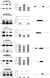
Endogenous target genes of YT521-B. RT-PCR analysis of YT521-B-dependent exons. Exons were identified by Splicearray analysis. High scoring events were analyzed by RT-PCR using primers in the flanking exons. YT521-B: overexpression of EGFP-YT521-B, YTHdel: overexpression of EGFP-YT521-Bdel(YTH). The image on the right shows a representative RT-PCR analysis, the graph in the middle shows the statistical evaluation of each experiment, and the schematic on the left shows the localization of YTH binding signatures on top of the annotated transcript structure. The processing of GSK3B was shown to be YT521-B independent both by Splicearray analysis and by RT-PCR.

References

    1. Chen Y., Varani G. (2005) FEBS J. 272, 2088–2097 - PubMed
    1. Maris C., Dominguez C., Allain F. H. (2005) FEBS J. 272, 2118–2131 - PubMed
    1. Pérez-Arellano I., Gallego J., Cervera J. (2007) FEBS J. 274, 4972–4984 - PubMed
    1. Theobald D. L., Mitton-Fry R. M., Wuttke D. S. (2003) Annu. Rev. Biophys. Biomol. Struct. 32, 115–133 - PMC - PubMed
    1. Lie Y. S., Macdonald P. M. (1999) Development 126, 1129–1138 - PubMed (VSports app下载)

Publication types (V体育2025版)

MeSH terms