Skip to main page content
U.S. flag

An official website of the United States government

Dot gov

The . gov means it’s official. Federal government websites often end in . gov or . mil VSports app下载. Before sharing sensitive information, make sure you’re on a federal government site. .

Https

The site is secure V体育官网. The https:// ensures that you are connecting to the official website and that any information you provide is encrypted and transmitted securely. .

. 2009 Dec 15;8(24):4168-75.
doi: 10.4161/cc.8.24.10800.

The p53 target Plk2 interacts with TSC proteins impacting mTOR signaling, tumor growth and chemosensitivity under hypoxic conditions

Affiliations

The p53 target Plk2 interacts with TSC proteins impacting mTOR signaling, tumor growth and chemosensitivity under hypoxic conditions

Elizabeth M Matthew et al. Cell Cycle. .

Abstract

Tuberous sclerosis complex 1 (TSC1) inhibits mammalian target of rapamycin (mTOR), a central promotor of cell growth and proliferation. The protein product of the TSC1 gene, hamartin (referred to as TSC1) is known to interact with Polo-like kinase 1 (Plk1) in a cell cycle regulated, phosphorylation-dependent manner. We hypothesized that the p53 target gene, Plk2, is a tumor suppressor, mediating its tumor suppressor function through interactions with TSC1 that facilitate TSC1/2 restraint of mTOR under hypoxic stress. We found that human lung tumor cells deficient in Plk2 grew larger than control tumors, and that Plk2 interacts with endogenous TSC1 protein. Additionally, C-terminal Plk2-GST fusion protein bound both TSC1 and TSC2 proteins. TSC1 levels were elevated in response to Adriamycin and cells transiently overexpressing Plk2 demonstrated decreased phosphorylation of the downstream target of mTOR, ribosomal protein p70S6 kinase during hypoxia. Plk2 levels were inversely correlated with cytoplasmic p70S6K phosphorylation. Plk2 levels did not increase in response to DNA damage (Adriamycin, CPT -11) when HCT 116 and H460 cells were exposed to hypoxia. TSC1-deficient mouse embryonic fibroblasts with TSC1 added back demonstrated decreased S6K phosphorylation, which was further decreased when Plk2 was transiently overexpressed. Interestingly, under normoxia, Plk2 deficient tumor cells demonstrated increased apoptosis in response to various chemotherapeutic agents including CPT -11 but increased resistance to apoptotic death after CPT-11 treatment under hypoxia, and tumor xenografts comprised of these Plk2-deficient cells were resistant to CPT -11. Our results point to a novel Plk2-TSC1 interaction with effects on mTOR signaling during hypoxia, and tumor growth that may enable targeting Plk2 signaling in cancer therapy. VSports手机版.


"VSports在线直播" Figures
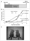
Figure 1
Figure 1
Plk2-deficiency stimulates tumor growth. (A) Mice were inoculated with Plk2 deficient (or control) H460 non-small cell lung carcinoma cells. One week after inoculation, mice were treated with 40 mg/kg camptothecin, three times per week (or untreated) and sacrificed at day 20 post-inoculation. (B) Representative growth curves. (C) Representative mice with control (left flank of mouse) and Plk2 deficient (right flank of mouse) tumors at day 14.
Figure 2
Figure 2
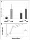
Plk2-deficiency stimulates tumor growth and camptothecin resistance in vivo not in vitro. (A) Mice bearing Plk2 deficient H460 non-small cell lung carcinoma cell xenografts (Fig. 1) were measured for tumor volume at day 20. (B) Two H460 Plk2 knock-down clones (Snk9; Snk7) and control (pSuper1) were plated at equal densities and counted every 24 hours for 7 days. Cell numbers were plotted on a log scale.
Figure 3
Figure 3
p53 and mTOR pathways cross-talk: Plk2 interacts with TSC1 and inhibits down-stream mTOR signaling and DNA damage stimulates TSC1 expression. (A) Plk2-TSC1 interaction. H460 cell lysates were precleared, half the cell lysate was incubated with Anti-TSC1 antibody and the other half with equal μgrams of whole mouse IgG. Protein-antibody complexes were immunoprecipitated with protein agarose A/G beads, run on an 11% SDS-PAGE gel and immunoblotted for TSC1 and Plk2. (B) DNA Damage: TSC1. HCT 116 cells were treated with Adriamycin (0.2 μg/mL, 20 hours). Cell lysates were run on an 7.5% SDS-PAGE gel and immunoblotted for hamartin (TSC1), phosphorylated p70S6K and β-actin (loading control). (C) DNA Damage: TSC1 HCT 116 cells were untreated or treated with 0.2 μg/mL of Adriamycin for 10 hours. Cell lysates were run on an SDS-PAGE gel and immunoblotted for hamartin (TSC1) and β-actin (loading control). (D) Hypoxia: Plk2 overexpression and mTOR signaling. HCT 116 cells were transfected with pcDNA3.1V5His-Plk2 (Plk2) or pcDNAV5His (Control = Cntrl). Forty hours post-transfection, cells were placed in hypoxia (0.2% oxygen) for 12 hours, harvested and the lysates run on a 4–12% Bis-Tris gel and immunoblotted for V5, phospho-p70 S6K and β-actin.
Figure 4
Figure 4
Plk2 interactions with TSC1 and TSC2 are cell-type independent. pGEX4T3 Empty (GST) or pGEX4T3 expressing Carboxy-Terminal Plk2 (CT-Plk2 GST) were run over glutathione-Sepharose beads followed by PBS wash (2x), untreated H460 (A and B), HCT 116 (C) or T3 MEF (D and E) cell lysates and PBS wash (2x). Glutathione elution buffer was used to elute or the beads were harvested directly. Equal volume Laemmli was added to eluate or beads, boiled and loaded in duplicate with input onto an SDS-PAGE gel. After transfer and blocking (5% NFDM), membranes were probed with TSC1, TSC2 and Plk2.
Figure 5
Figure 5
Plk2 facilitates TSC1 inhibition of mTOR: decreased phosphorylation of p70S6K. TSC1−/− MEFs with empty vector (P2) or hTSC1 stably re-introduced (T3) were transiently transfected with pcD-NAV5His (V) or pcDNAV5His-Plk2 (S). Forty hours post-transfection, cells were kept in normoxia or exposed to hypoxia (0.5%, 8 hrs), harvested on ice, washed with PBS and lysed in PTY buffer with protease and phosphatase inhibitors. Cell lysates were mixed with equal volume 2x sample buffer, boiled and loaded on an 11% SDS-PAGE gel.
Figure 6
Figure 6
Plk2 levels are not increased by DNA damage under hypoxia. (A) H460 cells were treated with CPT-11 (100 μg/mL) for 22 hours. After the first 2.5 hours of CPT-11, cells were placed in 0.2% hypoxia for 19.5 hrs, harvested, run on a 7.5% SDS-PAGE gel and immunoblotted for Plk2 and Ran (loading control). (B) HCT 116 cells were treated with Adriamycin (0.2 μg/mL, 20 hrs). Cell lysates were run on a 7.5% SDS-PAGE gel and immunoblotted for Plk2 and β-actin (loading control).
Figure 7
Figure 7
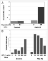
Plk2-deficient cells demonstrated increased sensitivity to DNA damage in vitro under normoxic conditions. H460 cells with Plk2 stably knocked-down were treated with either (A) Etoposide (4 mM, 34 hrs) or (B) Camptothecin (24 hrs) at increasing concentrations. The percentage of cells with sub-G1 DNA content was determined through propidium iodide staining and flow cytometry. Control, pSuper empty vector; Plk2 KD, Plk2 Knock Down.
Figure 8
Figure 8
Plk2-deficient cells are chemoresistant under hypoxia and chemosensitive under normoxia in vitro. Plk2 deficient or control H460 cells were treated with camptothecin (0, 100 or 200 μg; 22 hrs includes 2.5 hrs pre-hypoxia) and hypoxia (0.2% oxygen, 19 hrs) or normoxia. Cells were harvested, fixed and stained with propidium iodide and analyzed by flow cytometry for sub-G1 DNA content.
Figure 9
Figure 9
Schematic model for regulation of mTOR pathway signaling through interaction of the p53 target Plk2 with TSC proteins. Our model proposes that Plk2 and TSC1 interact to facilitate TSC1/TSC2 restraint of mTOR. Decreased mTOR activity leads to decreased phosphorylation of p70S6K and consequent decrease in protein translation, cell growth and proliferation. These events are regulated by hypoxia and DNA damage.

References (V体育官网)

    1. Guertin D, Sabatini DM. Defining the role of mTOR in cancer. Cancer Cell. 2007;12:9–22. - PubMed
    1. Crino PB, Hathanson KL, Henske EP. The tuberous sclerosis complex. N Engl J Med. 2006;355:1345–56. - PubMed
    1. Astrinidis A, Senapedis W, Henske EP. Hamartin, the tuberous sclerosis complex 1 gene product, interacts with polo-like kinase 1 in a phosphorylation-dependent manner. Human Mol Genet. 2006;15:287–97. - PubMed
    1. Burns TF, Fei P, Scata KA, Dicker DT, El-Deiry WS. Silencing of the novel p53 target gene Snk/Plk2 leads to mitotic catastrophe in paclitaxel (taxol)-exposed cells. Mol Cell Biol. 2003;23:5556–71. - PMC - PubMed
    1. Beuvink I, Boulay A, Fumagalli S, Zilbermann F, Reutz S, O’Reilly T, et al. The mTOR inhibitor RAD001 sensitizes tumor cells to DNA-damage induced apoptosis through inhibition of p21 translation. Cell. 2005;120:747–59. - PubMed

Publication types

MeSH terms

V体育ios版 - Substances