Skip to main page content
U.S. flag

An official website of the United States government

Dot gov

The . gov means it’s official. Federal government websites often end in . gov or . mil. Before sharing sensitive information, make sure you’re on a federal government site. VSports app下载.

Https

The site is secure V体育官网. The https:// ensures that you are connecting to the official website and that any information you provide is encrypted and transmitted securely. .

. 2010 Mar;101(3):658-65.
doi: 10.1111/j.1349-7006.2009.01427.x. Epub 2009 Nov 7.

"V体育官网入口" Myristoylated alanine-rich C kinase substrate phosphorylation promotes cholangiocarcinoma cell migration and metastasis via the protein kinase C-dependent pathway

Affiliations

Myristoylated alanine-rich C kinase substrate phosphorylation promotes cholangiocarcinoma cell migration and metastasis via the protein kinase C-dependent pathway

V体育2025版 - Anchalee Techasen et al. Cancer Sci. 2010 Mar.

Abstract

Myristoylated alanine-rich C kinase substrate (MARCKS), a substrate of protein kinase C (PKC) has been suggested to be implicated in cell adhesion, secretion, and motility through the regulation of the actin cytoskeletal structure. The quantitative real-time-polymerase chain reaction analysis revealed that MARCKS is significantly overexpressed in Opisthorchis viverrini-associated cholangiocarcinoma (CCA) (P = 0. 001) in a hamster model, which correlated with the results of mRNA in situ hybridization. An immunohistochemical analysis of 60 CCA patients revealed a significant increase of MARCKS expression. Moreover, the log-rank analysis indicated that CCA patients with a high MARCKS expression have significantly shorter survival times than those with a low MARCKS expression (P = 0. 02). This study investigated whether MARCKS overexpression is associated with CCA metastasis. Using a confocal microscopic analysis of CCA cell lines that had been stimulated with the PKC activator, 12-0-tetradecanoyl phorbol-13-acetate (TPA), MARCKS was found to be translocated from the plasma membrane to the perinuclear area. In addition, phosphorylated MARCKS (pMARCKS) became highly concentrated in the perinuclear area VSports手机版. Moreover, an adhesion assay demonstrated that the exogenous overexpression of MARCKS remarkably promoted cell attachment. Interestingly, after TPA stimulation, the CCA cell line-depleted MARCKS showed a decrease in migration and invasion activity. It can be concluded that in non-stimulation, MARCKS promotes cell attachment to the extracellular matrix. After TPA stimulation, PKC phosphorylates MARCKS leading to cell migration or invasion. Taken together, the results of this study reveal a prominent role for MARCKS as one of the key players in the migration of CCA cells and suggest that cycling between MARCKS and pMARCKS can regulate the metastasis of biliary cancer cells. .


Figures (V体育安卓版)

Figure 1
Figure 1
Quantitative real‐time–polymerase chain reaction assay was performed to determine the myristoylated alanine‐rich C kinase substrate (MARCKS) mRNA level in the tissues at different times after treatment with either Opisthorchis viverrini, N‐nitrosodimethylamine (NDMA) or both. Data are shown as mean ± SD. Asterisks denote significant increases in the MARCKS expression compared with the control. *P <0.05, **P =0.001.
Figure 2
Figure 2
Non‐radioactive in situ hybridization was used to determine myristoylated alanine‐rich C kinase substrate mRNA localization in the liver tissue. To verify the specificity of the cRNA probe, a hybridization mixture that contained a non‐digoxiginin (DIG)‐labeled antisense probe and a DIG‐labeled probe at a ratio of 30:1 was used as the negative control. (a) Non‐treated; (b) Opisthorchis viverrini treated; (c) N‐nitrosodimethylamine (NDMA) treated; (d) cholangiocarcinoma (CCA); Ov plus NDMA‐treated groups at (e) week 1; (f) week 2; (g) week 3; (h) negative control.
Figure 3
Figure 3
Immunohistochemistry of myristoylated alanine‐rich C kinase substrate (MARCKS) (a) and phosphorylated MARCKS (pMARCKS) (c) in cholangiocarcinoma (CCA) tissues. Poorly (i), moderately (ii), papillary (iii), and well‐differentiated (iv) CCA showed a strong expression of MARCKS. Some CCA cells in the bile duct showed a strong intensity of pMARCKS. Original magnification ×40. Survival curves calculated for MARCKS (b) and pMARCKS (d) according to the Kaplan–Meier log–rank test (P =0.02 and P =0.12, respectively). (––), high level of MARCKS or pMARCKS, (‐ ‐ ‐), low level of MARCKS or pMARCKS.
Figure 4
Figure 4
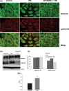
Intracellular localization of myristoylated alanine‐rich C kinase substrate (MARCKS) and phosphorylated MARCKS (pMARCKS) (a). Cholangiocarcinoma (CCA) cells were stimulated with 10 ng/mL 12‐0‐tetradecanoyl phorbol‐13‐acetate (TPA) or 5 μm GF109203x before TPA treatment. Confocal laser microscopy with an objective lens of ×40 was employed. In the control group, MARCKS was mostly located at the plasma membrane and pMARCKS was evenly distributed throughout the cytoplasm. Upon TPA treatment, MARCKS was translocated to the cytoplasm and the perinuclear area, and pMARCKS became highly detectable at the perinuclear level. Upon GF109203x + TPA treatment, MARCKS and pMARCKS distribution and abundance were similar to those observed in non‐stimulated CCA cells. Western blotting was used for protein checking (b). Apparent intensity of bands on the membranes was estimated using a densitometer (c). Protein kinase C (PKC) activities were compared between the control and TPA‐treated cells (d). Asterisk denotes a significant increase of PKC activity when compared with the control. *P <0.05.
Figure 5
Figure 5
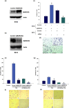
Gain‐on and gain‐off function of myristoylated alanine‐rich C kinase substrate (MARCKS). Western blot analysis was performed to confirm the overexpression of MARCKS in the M156 cholangiocarcinoma (CCA) cell line in comparison to the empty vector control cells (a) and RNAi against MARCKS in the M214 CCA cell line (b). MARCKS promotes cell re‐attachment in CCA cell lines (c,d). Re‐attachment assay was employed. Values are expressed as mean ± SD. Asterisk denotes a significant increase. *P <0.05. In contrast, the knocking down of endogenous MARCKS in the M214 CCA cell line leads to a decrease in cell migration or invasion after 12‐0‐tetradecanoyl phorbol‐13‐acetate (TPA) stimulation. Migration (e,f) or invasion (g,h) assays were performed in TPA‐treated control cells (i), under TPA‐treated siRNA conditions (ii), GF109203x with TPA‐treated control cells (iii), and GF109203x under TPA‐treated siRNA conditions (iv). Values are expressed as mean ± SD. Asterisk denotes a significant decrease. *P <0.05.
Figure 6
Figure 6
Intracellular localization of myristoylated alanine‐rich C kinase substrate (MARCKS) and actin filaments (F‐actin). Cholangiocarcinoma cells were stimulated with 10 ng/mL 12‐0‐tetradecanoyl phorbol‐13‐acetate (TPA) or 5 μm GF109203x before TPA treatment. Immunofluorescence microscopy with an objective lens of ×40 was employed. In the control group, MARCKS and F‐actin were colocalized at the plasma membrane. Upon TPA treatment, MARCKS was translocated to cytoplasm at the perinuclear area, and F‐actin disassembly was found. In GF109203x + TPA treatment, MARCKS and F‐actin distribution was similar to that observed in the control cells.

References

    1. Elkins DB, Haswell‐Elkins MR, Mairiang E et al. A high frequency of hepatobiliary disease and suspected cholangiocarcinoma associated with heavy Opisthorchis viverrini infection in a small community in north‐east Thailand. Trans R Soc Trop Med Hyg 1990; 84 (5): 715–9. - PubMed
    1. Elkins DB, Mairiang E, Sithithaworn P et al. Cross‐sectional patterns of hepatobiliary abnormalities and possible precursor conditions of cholangiocarcinoma associated with Opisthorchis viverrini infection in humans. Am J Trop Med Hyg 1996; 55 (3): 295–301. - PubMed
    1. Haswell‐Elkins MR, Mairiang E, Mairiang P et al. Cross‐sectional study of Opisthorchis viverrini infection and cholangiocarcinoma in communities within a high‐risk area in northeast Thailand. Int J Cancer 1994; 59 (4): 505–9. - VSports - PubMed
    1. Flavell DJ, Lucas SB. Promotion of N‐nitrosodimethylamine‐initiated bile duct carcinogenesis in the hamster by the human liver fluke, Opisthorchis viverrini . Carcinogenesis 1983; 4 (7): 927–30. - PubMed
    1. Ohshima H, Bartsch H. Chronic infections and inflammatory processes as cancer risk factors: possible role of nitric oxide in carcinogenesis. Mutat Res 1994; 305 (2): 253–64. - PubMed

"VSports" Publication types

"V体育官网入口" MeSH terms