<acronym id="6E1du"></acronym>
Skip to main page content (VSports app下载)
U.S. flag

An official website of the United States government

Dot gov

The . gov means it’s official VSports app下载. Federal government websites often end in . gov or . mil. Before sharing sensitive information, make sure you’re on a federal government site. .

Https

The site is secure. The https:// ensures that you are connecting to the official website and that any information you provide is encrypted and transmitted securely V体育官网. .

. 2009 Dec 8;16(6):463-74.
doi: 10.1016/j.ccr.2009.10.016.

VSports最新版本 - Somatic mutations in p85alpha promote tumorigenesis through class IA PI3K activation

Affiliations

"VSports app下载" Somatic mutations in p85alpha promote tumorigenesis through class IA PI3K activation

Bijay S Jaiswal et al. Cancer Cell. .

Abstract

Members of the mammalian phosphoinositide-3-OH kinase (PI3K) family of proteins are critical regulators of various cellular process including cell survival, growth, proliferation, and motility. Oncogenic activating mutations in the p110alpha catalytic subunit of the heterodimeric p110/p85 PI3K enzyme are frequent in human cancers. Here we show the presence of frequent mutations in p85alpha in colon cancer, a majority of which occurs in the inter-Src homology-2 (iSH2) domain. These mutations uncouple and retain p85alpha's p110-stabilizing activity, while abrogating its p110-inhibitory activity. The p85alpha mutants promote cell survival, AKT activation, anchorage-independent cell growth, and oncogenesis in a p110-dependent manner. VSports手机版.


Figures
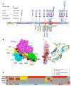
Figure 1
Figure 1. p85α mutations in human cancer
(A) Cartoon depicting the p85α somatic mutations. Horizontal bar below mutated residues indicates conserved residues across the p85 family members (R1-R3). Black and orange vertical bars indicate previously identified p85α mutations (Jimenez et al., 1998; Jucker et al., 2002; Parsons et al., 2008; Philp et al., 2001; TCGA, 2008; Wood et al., 2007). Orange bars signify hotspots, with the bar height drawn proportional to number of times the residue/region has been altered. (B) p85α somatic mutations modeled on a recent crystal structure (PDB code 2RD0) of p85α iSH2 domain in complex with p110α (Huang et al., 2007). Blue patches on the surface of p110α represent regions that are <3.5Å from p85α iSH2. (C) The p85α residues, N564 and D560 within hydrogen bonding distance of N345 of p110α (Huang et al., 2008; TCGA, 2008) located in the C2 domain is shown. Models of p110β (yellow) and p110δ (purple) C2 domains superimposed on p110α C2 domain (cyan) illustrates the proximity of conserved residue N344 of p110β and N334 of p110δ (corresponding to N345 of p110α) and p85α iSH2 residues D560 and N564. (D) Mutation map of tumors carrying changes in one or more of the genes studied. Each column corresponds to a tumor sample. Pc = pancreatic cancer. 1Nature 455, 1061, 2Science 321, p1807, 3Science 318, p1108 and 4The EMBO journal 17, p743.
Figure 2
Figure 2. p85α mutants interact with p110α and stabilize p110α
(A-D) HA-tagged p85α mutants and wild-type p85α that carry an intact p110α interaction domain immunoprecipitate p110α from Cos7 cells transfected with the constructs indicated in the panels. Expression of all the transfected constructs was confirmed in the lysates as shown below in each of the panels. The dominant negative p85Δ and empty vector transfected cells served as controls. (E) p85α mutants that contain an intact p110α binding domain when transiently expressed in pan-p85 null MEFs stabilize p110α levels.
Figure 3
Figure 3. p85α mutants interact with p110β and p110δ
(A-B) Indicated HA-tagged p85α mutants and wild-type p85α co-transfected with myc-p110β (A) or myc-p110δ (B) immunoprecipitated p110β (A) or p110δ (B). Expression of all the transfected constructs was confirmed in the lysates. The dominant negative p85Δ and empty vector transfected cells served as controls.
Figure 4
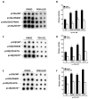
Figure 4. p85α mutants are impaired in regulating class IA PI3K activity
(A, C, E) Dot blots show the amount of phosphorylated lipid generated by each of the tested heterodimeric enzymes in triplicates. (B, D, F) Mean ± SEM of the amount of phosphorylated lipid generated in (A, C, E) by each of the enzyme complexes in arbitrary units. Elevated PI3K activity is effectively inhibited by a p110α/δ (B, F) or p110β (TGX-221; D) inhibitor (Folkes et al., 2007; Jackson et al., 2005). Mean ± SEM % control (p85α-WT) activity is shown. (*p<0.05, **p<0.01 and ***p<0.001)
Figure 5
Figure 5. p85α mutants promote p110-dependent cell survival and Akt signaling
(A-H) Stable expression p85α mutants alone (A, E), together with p110α (B, F) or p110β (C, G) or p110δ (D, H) promote IL3-independent survival of BaF3 cells (E-H) and elevated pAkt (A-D) levels. The cell survival effects (E-H) of p85α mutants are inhibited by a p110α/δ or p110β (TGX-221) inhibitor (Folkes et al., 2007; Jackson et al., 2005). Data shown in (E-H) are mean ± SEM of at least three independent experiments with multiple replicates. (*p<0.05 and **p<0.01)
Figure 6
Figure 6. p85α mutants promote anchorage independent growth, p110 stabilization and proliferation
(A-F) A representative image showing anchorage-independent growth (A-C) and number of colonies formed in the absence or presence of PI3K inhibitors (D-F) by BaF3 cells stably expressing p85α mutants together with p110α (A), p110β (B) or p110δ (C). (G, H) Pan-p85 null MEFs stably reconstituted with p85α mutants show stabilization of p110α̣ p110β and p110δ (G), elevated pAkt (G) and increased cell proliferation (H). Data shown (D-F, H) are mean ± SEM. (*p<0.05 and **p<0.01).
Figure 7
Figure 7. p85α mutants lead to reduced overall survival
(A) Kaplan-Meier survival curves for cohorts of mice implanted with p85α mutant expressing BaF3 cells compared to vector control cells or p85α wild-type cells or p110α cells alone or p85α /p110α cells showed a significant reduction in overall survival (n = 10 for arms; Log-rank test p<0.0001). (B) Flow cytometric analysis of total bone marrow isolated from mice receiving GFP- and/or dsRed-tagged BaF3 cells expressing the various p85α mutants. (C) Mean number of GFP and/or dsRed positive cells in the bone marrow of mice (n = 3) that received BaF3 cells expressing p85α mutants. Values are mean ± SEM. (D) Representative H&E-stained liver (top) and spleen (bottom) sections from the same mice analyzed in (B). Arrows indicate tumor cells infiltrating the liver. R = red pulp, W = lymphoid follicles of white pulp. In unmarked spleen section, there is a loss of red/white pulp architecture due to disruption by infiltrating tumor cells. Scale bar corresponds to 100μm.

Comment in

References

    1. Amzel LM, Huang CH, Mandelker D, Lengauer C, Gabelli SB, Vogelstein B. Structural comparisons of class I phosphoinositide 3-kinases. Nat Rev Cancer. 2008;8:665–669. - PMC - PubMed
    1. Backer JM, Myers MG, Jr., Shoelson SE, Chin DJ, Sun XJ, Miralpeix M, Hu P, Margolis B, Skolnik EY, Schlessinger J, et al. Phosphatidylinositol 3′-kinase is activated by association with IRS-1 during insulin stimulation. Embo J. 1992;11:3469–3479. - "V体育2025版" PMC - PubMed
    1. Bader AG, Kang S, Zhao L, Vogt PK. Oncogenic PI3K deregulates transcription and translation. Nat Rev Cancer. 2005;5:921–929. - PubMed
    1. Borlado LR, Redondo C, Alvarez B, Jimenez C, Criado LM, Flores J, Marcos MA, Martinez AC, Balomenos D, Carrera AC. Increased phosphoinositide 3-kinase activity induces a lymphoproliferative disorder and contributes to tumor generation in vivo. Faseb J. 2000;14:895–903. - PubMed
    1. Brachmann SM, Yballe CM, Innocenti M, Deane JA, Fruman DA, Thomas SM, Cantley LC. Role of phosphoinositide 3-kinase regulatory isoforms in development and actin rearrangement. Mol Cell Biol. 2005;25:2593–2606. - PMC - PubMed

Publication types

Substances (V体育平台登录)

Associated data