Skip to main page content
U.S. flag

An official website of the United States government

Dot gov

The . gov means it’s official. Federal government websites often end in VSports app下载. gov or . mil. Before sharing sensitive information, make sure you’re on a federal government site. .

Https

The site is secure. The https:// ensures that you are connecting to the official website and that any information you provide is encrypted and transmitted securely. V体育官网.

. 2009 Dec;10(12):1785-801.
doi: 10.1111/j.1600-0854.2009.00994.x. Epub 2009 Oct 5.

Hydrophobic and basic domains target proteins to lipid droplets

Affiliations

"VSports" Hydrophobic and basic domains target proteins to lipid droplets

Mercedes Ingelmo-Torres (VSports) et al. Traffic. 2009 Dec.

Abstract (V体育官网)

In recent years, progress in the study of the lateral organization of the plasma membrane has led to the proposal that mammalian cells use two different organelles to store lipids: intracellular lipid droplets (LDs) and plasma membrane caveolae. Experimental evidence suggests that caveolin (CAV) may act as a sensitive lipid-organizing molecule that physically connects these two lipid-storing organelles. Here, we determine the sequences necessary for efficient sorting of CAV to LDs. We show that targeting is a process cooperatively mediated by two motifs. CAV's central hydrophobic domain (Hyd) anchors CAV to the endoplasmic reticulum (ER). Next, positively charged sequences (Pos-Seqs) mediate sorting of CAVs into LDs. Our findings were confirmed by identifying an equivalent, non-conserved but functionally interchangeable Pos-Seq in ALDI, a bona fide LD-resident protein. Using this information, we were able to retarget a cytosolic protein and convert it to an LD-resident protein. Further studies suggest three requirements for targeting via this mechanism: the positive charge of the Pos-Seq, physical proximity between Pos-Seq and Hyd and a precise spatial orientation between both motifs. The study uncovers remarkable similarities with the signals that target proteins to the membrane of mitochondria and peroxisomes VSports手机版. .


"VSports注册入口" Figures
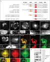
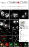
Figure 1
Figure 1. Identification of the LD-targeting motifs contained in CAVs
A) Schematic representation of the mutants of CAV3 and CAV2 used in this figure. The Hyd has been colored in red and the CSD in orange. Numbers on the residues indicate the relative position of each residue within the full-length protein with respect to the N-terminal end (see in addition Table 1). Letters between brackets indicate the corresponding panel for each mutant. Mutants were N-terminally tagged with GFP, expressed in lipid-loaded cells and their final distribution was analyzed by fluorescence microscopy (B–J). I) Shows the colocalization between CAV3LLS (green) and Nile Red-positive LDs (red). J) Shows the colocalization of CAV3LLS-deltaEnd (green) and the endogenous ER-resident protein, Sec61 (red). Arrows mark the selected areas for the high magnification panels. Scale bar is 20 μm. K) Distribution of the mutants in sucrose density gradients. Cells were treated as indicated above and additionally incubated for 12 h with cycloheximide. Fractions were collected from the top of the gradient and the distribution of the mutants (detected by means of western blotting and anti-GFP antibodies) was compared with GFP, Sec61 and GFP-ADRP. `LDs' is the top fraction of the gradient in which LDs fractionate and `b' corresponds to the bottom fraction of the gradient. See text and tables for more details.
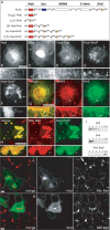
Figure 2
Figure 2. Analysis of the Pos-Seqs in full-length CAVs
A) Schematic representation of mutants analyzed in the figure. Letters between brackets indicate the corresponding panel for each mutant. Numbers on the residues indicate the relative position of each residue within the full-length protein with respect to the N-terminal end (see in addition Table 1). The Hyd has been colored in red. Mutants were N-terminally tagged with GFP, expressed in lipid-loaded cells for 24 h, additionally treated with cycloheximide for additional 6 h and their final distribution was analyzed by fluorescence microscopy (B–F). Scale bar is 20 μm. G and H) Distribution of the mutants in sucrose density gradients as in Figure 1K. The distribution of the mutants along the gradients was quantified as the relative intensity of each band with respect to the total intensity of the six bands. Endogenously expressed ADRP and Sec61 were used to define the relative position of LD and the ER membranes within the gradients. I and J) Low magnification panels to show LD formation (red) in cells transfected with CAV3 or CAV36+ to g (in green). Discontinuous lines indicate the edge of transfected cells. K and L) Colocalization of CAV36+ to g (green) with the endogenous ER-resident protein, Sec61 (red in K) and the endogenous Golgi marker GM130 (red in L). Scale bar is 20 μm.
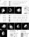
Figure 3
Figure 3. Identification of the LD-targeting motifs contained in ALDI
A) Schematic representation of the mutants used in the figure. The Hyd has been colored in red and the juxtamembrane region (Jux) in blue. Numbers on the residues indicate the relative position of each residue within the full-length protein with respect to the N-terminal end (see in addition Table 1). Letters between brackets indicate the corresponding panel for each mutant. Mutants were N-terminally tagged with GFP, expressed in lipid-loaded cells and their final distribution was analyzed by fluorescence microscopy (B–G). G) Shows the colocalization between ALDI-deltaEnd (green) and the ER-resident protein, Sec61 (red). Arrows mark the selected areas for high magnification. Scale bar is 20 μm. H) Distribution of the mutants in sucrose density gradients as in Figure 1K.
Figure 4
Figure 4. The Hyd and the Pos-Seq cooperate for targeting to LDs
A) Schematic representation of the mutants used in this figure. The three studied sequences have been colored: Hyd (red), the Jux (blue) and the End (orange). Numbers on the residues indicate the relative position of each residue within the full-length protein with respect to the N-terminal end (see in addition Table 1). Letters between brackets indicate the corresponding panel for each mutant. Mutants were N-terminally tagged with GFP, expressed in lipid-loaded cells and their final distribution was analyzed by fluorescence microscopy (B–H). Arrows mark the selected areas for high magnification (G and H). Shows the colocalization between the Hyd (green) and endogenous Sec61 (red) and the Hyd-End3 (green) with Nile Red (red). I) Distribution of the mutants in sucrose density gradients as described in Figure 1. J and K) Low magnification panels to illustrate that cells transfected as above with Hyd and with ER (in green) display a normal formation of LDs (in red), although the mutants accumulate in the ER or the cytosol, respectively. Discontinuous lines indicate the edge of transfected cells. Scale bar is 20 μm.
Figure 5
Figure 5. Spatial determinants for sorting the model protein to LDs
A) Schematic representation of mutants analyzed in the figure. Negative numbers on the residues indicate the relative position with respect to the C-terminal end. Letters between brackets indicate the corresponding panel for each mutant. The Hyd has been colored in gray. The residues that separate the Hyd and the positives charges of the Jux-Seq are underlined. Mutants were N-terminally tagged with GFP, expressed in lipid-loaded cells and their final distribution was analyzed by fluorescence microscopy (B–D). E) Distribution of the mutants in sucrose density gradients. F) Schematic representation of the proteins analyzed in this figure. Negative numbers on the residues indicate the relative position with respect to the C-terminal end. The Hyd has been colored in gray. Letters between brackets indicate the corresponding panel for each mutant. All the constructs were N-terminally tagged with GFP, expressed in lipid-loaded cells and their final distribution was analyzed by fluorescence microscopy (G–K). Arrows mark the selected areas for the high magnification panels. l) Distribution of the mutants treated as in Figure 1 in sucrose density gradients. M) Mutants were transfected for 12 h and then fractionated in sucrose density gradients. Scale bar is 20 μm.
Figure 6
Figure 6. Differential sorting of Hyd and Hyd-End3 within the ER
GFP-Hyd and orange-Hyd-End3 were cotransfected in starved cells and cells were selected under the microscope for coexpression of both proteins (A). Next, fatty acids were added to the cells and images were captured every 2 min. B) The figure shows the selected image 4 h after the treatment with fatty acids and arrows mark the selected areas for high magnification. C) The similar expression levels of the mutants were confirmed by western blotting. D) Cotransfected GFP-Hyd (green) and orange-Hyd-End3 (red) in PFA-fixed starved cells. Scale bar is 20 μm.

References

    1. Murphy S, Martin S, Parton RG. Lipid droplet-organelle interactions; sharing the fats. Biochim Biophys Acta. 2009;1791:441–447. - PubMed
    1. Ohsaki Y, Cheng J, Suzuki M, Shinohara Y, Fujita A, Fujimoto T. Biogenesis of cytoplasmic lipid droplets: from the lipid ester globule in the membrane to the visible structure. Biochim Biophys Acta. 2009;1791:399–407. - VSports - PubMed
    1. Tauchi-Sato K, Ozeki S, Houjou T, Taguchi R, Fujimoto T. The surface of lipid droplets is a phospholipid monolayer with a unique fatty acid composition. J Biol Chem. 2002;277:44507–44512. - VSports app下载 - PubMed
    1. Leber R, Zinser E, Zellnig G, Paltauf F, Daum G. Characterization of lipid particles of the yeast, Saccharomyces cerevisiae. Yeast. 1994;10:1421–1428. - PubMed
    1. Bartz R, Li WH, Venables B, Zehmer JK, Roth MR, Welti R, Anderson RG, Liu P, Chapman KD. Lipidomics reveals that adiposomes store ether lipids and mediate phospholipid traffic. J Lipid Res. 2007;48:837–847. - PubMed

Publication types

"VSports" LinkOut - more resources