Skip to main page content (V体育ios版)
U.S. flag

An official website of the United States government

Dot gov

The VSports app下载. gov means it’s official. Federal government websites often end in . gov or . mil. Before sharing sensitive information, make sure you’re on a federal government site. .

Https

The site is secure. The https:// ensures that you are connecting to the official website and that any information you provide is encrypted and transmitted securely. V体育官网.

. 2009 Sep 17;4(9):e7084.
doi: 10.1371/journal.pone.0007084.

SBDS expression and localization at the mitotic spindle in human myeloid progenitors (VSports在线直播)

Affiliations

SBDS expression and localization at the mitotic spindle in human myeloid progenitors

Claudia Orelio et al. PLoS One. .

Abstract

Background: Shwachman-Diamond Syndrome (SDS) is a hereditary disease caused by mutations in the SBDS gene. SDS is clinically characterized by pancreatic insufficiency, skeletal abnormalities and bone marrow dysfunction. The hematologic abnormalities include neutropenia, neutrophil chemotaxis defects, and an increased risk of developing Acute Myeloid Leukemia (AML). Although several studies have suggested that SBDS as a protein plays a role in ribosome processing/maturation, its impact on human neutrophil development and function remains to be clarified VSports手机版. .

Methodology/principal findings: We observed that SBDS RNA and protein are expressed in the human myeloid leukemia PLB-985 cell line and in human hematopoietic progenitor cells by quantitative RT-PCR and Western blot analysis. SBDS expression is downregulated during neutrophil differentiation. Additionally, we observed that the differentiation and proliferation capacity of SDS-patient bone marrow hematopoietic progenitor cells in a liquid differentiation system was reduced as compared to control cultures. Immunofluorescence analysis showed that SBDS co-localizes with the mitotic spindle and in vitro binding studies reveal a direct interaction of SBDS with microtubules V体育安卓版. In interphase cells a perinuclear enrichment of SBDS protein which co-localized with the microtubule organizing center (MTOC) was observed. Also, we observed that transiently expressed SDS patient-derived SBDS-K62 or SBDS-C84 mutant proteins could co-localize with the MTOC and mitotic spindle. .

Conclusions/significance: SBDS co-localizes with the mitotic spindle, suggesting a role for SBDS in the cell division process, which corresponds to the decreased proliferation capacity of SDS-patient bone marrow CD34(+) hematopoietic progenitor cells in our culture system and also to the neutropenia in SDS patients V体育ios版. A role in chromosome missegregation has not been clarified, since similar spatial and time-dependent localization is observed when patient-derived SBDS mutant proteins are studied. Thus, the increased risk of myeloid malignancy in SDS remains unexplained. .


"VSports app下载" Conflict of interest statement

Competing Interests: The authors have declared that no competing interests exist.

V体育ios版 - Figures

Figure 1
Figure 1. Generating SBDS Antibody.
To test specificity of our SBDS antibody we overexpressed GFP- or HA-tagged SBDS in HeLa or Cos-7 cells (A) Western blotting shows that the anti-HA antibody recognizes a 30–34 kD protein in HA-SBDS-FL transfected Cos-7 cells. In a duplicate blot we show that our SBDS antibody recognizes specifically a 30–34 kD protein in HA-SBDS transfected cells. Moreover, we detected an approximately 30 kD band, which represents the endogenous SBDS. (B) Immunostaining with our SBDS antibody shows that GFP-SBDS expression (green; top picture) and endogenous stained SBDS (red; middle picture) coincides in Cos-7 cells (bottom picture). (C) Schematic overview GFP-SBDS constructs used for transient transfection followed by Western blotting analysis. The grey, white and black regions indicate the three main SBDS protein regions, namely an N-terminal FYSH domain, a central helix-turn-helix motif and the C-terminal common fold which has homology with a RNA-Recognition Motif (RRM). (D) Western blot analysis for several GFP-SBDS protein isoforms transiently expressed in HeLa cells. GFP staining (top panel) shows that these constructs are expressed at expected molecular weight and staining with our SBDS antibody shows that the antibody recognizes GFP-SBDS-FL, R219, C84 and the N-terminally truncated SBDS isoforms (GFP-SBDS Δ1–65, Δ1–75 and Δ1–85), but not GFP-SBDS-K62. Protein standard indicates protein size as indicated (56 kD, 36 kD and 28 kD, respectively)
Figure 2
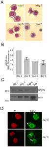
Figure 2. SBDS expression is downregulated during PLB-985 neutrophil differentiation.
(A) PLB-985 cells were cultured for several days in the presence of 0.5% DMF to induce differentiation. May-Grünwald-Giemsa staining of cytospins of differentiating PLB-985 cells. (B) Representative quantitative RT-PCR analysis shows that SBDS mRNA expression decreases 1.8 fold with PLB-985 neutrophil differentiation (n = 5), showing the mean±SEM. (C) Western blot analysis for SBDS expressing levels and actin as a protein loading control in undifferentiated (day 0) and differentiating (day 3–7) PLB-985 cells shows that SBDS protein expression decreases during neutrophil differentiation (representative for n = 5 independent cellular differentiation experiments). (D) Immunofluorescence staining for SBDS shows that SBDS localizes prominently to the nucleus (counterstained with propidium iodide) and to lower extent in the cytoplasm. A prominent perinuclear SBDS-enriched structure (indicated with the arrow head) was detected in PLB-985 cells.
Figure 3
Figure 3. SBDS expression is downregulated during human cord blood CD34+ neutrophil differentiation.
Human CD34+ cord blood cells were differentiated towards neutrophils with a cocktail of cytokines over a period of 18–21 days. (A) May-Grünwald-Giemsa staining on differentiating CD34+ cells shows that these cells are acquiring morphological characteristics of neutrophilic cells during the culture. (B) Representative quantitative RT-PCR analysis shows that SBDS mRNA expression decreases 5-fold with human cord blood neutrophil differentiation (n = 3; mean±SEM). (C) Immunofluorescence staining for SBDS in differentiating cord blood hematopoietic progenitor and neutrophilic cells shows that SBDS protein is localized predominantly in the nucleus and to a lower extent in the cytoplasm. In all cells a prominent SBDS-enrichment is observed in the perinuclear region (indicated with arrow). In mitotic cells, we observed that SBDS is localized at the mitotic spindle and/or centrosomes during the anaphase of mitosis (right panel).
Figure 4
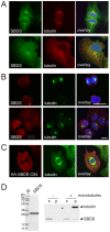
Figure 4. SBDS co-localizes with the MTOC and the mitotic spindle.
Endogenously expressed SBDS co-localizes with the mitotic spindle and/or centrosomes (top panel) and the MTOC area (bottom panel) in (A) HeLa cells (n = 8) and in (B) cord blood CD34+ cells (n = 3). (C) Overexpressed HA-SBDS-C84 localizes to the mitotic spindle in U2OS cells (n = 5). (D) Recombinant expressed SBDS was used as a ligand in a microtubule-binding protein spin-down assay. Right panel shows a Coomassie-stained gel with 5 µg of the purified SBDS protein with a molecular weight of 30 kD. Left panel shows a Coomassie- stained gel with SBDS protein incubated in the presence or absence of microtubules, as indicated by tubulin staining. In the absence of microtubules, the SBDS protein remains in the soluble fraction (s) and in the presence of 0.16 nM taxol-stabilized microtubules, a small, but significant fraction of the SBDS protein co-precipitates with microtubules in the pellet (p) fraction. Representative result of 5 independent experiments.
Figure 5
Figure 5. SBDS plays a role in myeloid progenitor proliferation.
BM CD34+ cells were differentiated towards neutrophils and monitored at several stages for proliferation and differentiation. (A) Average fold induction of cell numbers from 5 independent SDS and 3 independent control BM CD34+ differentiation cultures. Control BM cultures show an overall significantly higher fold induction in the cell number as compared to SDS patient cells. Fold induction was calculated within each experiment by dividing the number of cells in the culture at each time point by the number of cells that was seeded at day 0. Both for controls and SDS patients 0.15−0.5×10⁴ cells were seeded. Thereafter, the average fold was calculated. Error bars indicate SEM (Statistical analysis according to Mann Witney U testing, p = 0.014). (B) FACS analysis at day 17 shows that the percentage of CD11b+ cells in the SDS and control cultures is approximately 80% (control n = 3; SDS patient n = 2). (C) Representative May-Grunwald-Giemsa stained cells of control or SDS patient BM cultures at various days of the culture (control n = 3; SDS patient n = 5). (D) Bars indicate the average percentage of cells of the indicated differentiation status at day 17 of the culture. SDS BM differentiation cultures (dark bars) show less terminally differentiated neutrophils as compared to the control culture (light bars). However, the percentage of intermediately differentiated (myelocytic) cells and the percentage of macrophages was (slightly) increased in SDS patient cultures (n = 3 for control and n = 4 for SDS patient. Per experiment 196–232 cells were examined. 4×2, Chi-square test, p<0.03). May-Grünwald-Giemsa stained cells were morphologically analyzed and scored by two independent persons in a blind fashion.

References

    1. Shwachman H, Diamond LK, Oski FA, Khaw KT. The syndrome of pancreatic insufficiency and bone marrow dysfunction. J Pediatr. 1964;65:645–63. - PubMed
    1. Bodian M, Sheldon W, Lightwood R. Congenital hypoplasia of the exocrine pancreas. Acta Paediatr. 1964;53:282–93: 282-293. - PubMed
    1. Ginzberg H, Shin J, Ellis L, Morrison J, Ip W, et al. Shwachman syndrome: phenotypic manifestations of sibling sets and isolated cases in a large patient cohort are similar. J Pediatr. 1999;135:81–88. - PubMed
    1. Kuijpers TW, Alders M, Tool AT, Mellink C, Roos D, et al. Hematologic abnormalities in Shwachman Diamond syndrome: lack of genotype-phenotype relationship. Blood. 2005;106:356–361. - PubMed
    1. Shimamura A. Shwachman-Diamond Syndrome. Semin Hematol. 2006;43:178–188. - PubMed

Publication types

MeSH terms (V体育安卓版)