V体育ios版 - Skip to main page content
U.S. flag

An official website of the United States government

Dot gov

The . gov means it’s official. Federal government websites often end in . gov or VSports app下载. mil. Before sharing sensitive information, make sure you’re on a federal government site. .

Https

The site is secure. The https:// ensures that you are connecting to the official website and that any information you provide is encrypted and transmitted securely V体育官网. .

. 2009;11(4):R56.
doi: 10.1186/bcr2345. Epub 2009 Jul 30.

Breast-cancer-associated metastasis is significantly increased in a model of autoimmune arthritis

Affiliations

Breast-cancer-associated metastasis is significantly increased in a model of autoimmune arthritis

Lopamudra Das Roy et al. Breast Cancer Res. 2009.

Abstract (VSports)

Introduction: Sites of chronic inflammation are often associated with the establishment and growth of various malignancies including breast cancer. A common inflammatory condition in humans is autoimmune arthritis (AA) that causes inflammation and deformity of the joints VSports手机版. Other systemic effects associated with arthritis include increased cellular infiltration and inflammation of the lungs. Several studies have reported statistically significant risk ratios between AA and breast cancer. Despite this knowledge, available for a decade, it has never been questioned if the site of chronic inflammation linked to AA creates a milieu that attracts tumor cells to home and grow in the inflamed bones and lungs which are frequent sites of breast cancer metastasis. .

Methods: To determine if chronic inflammation induced by autoimmune arthritis contributes to increased breast cancer-associated metastasis, we generated mammary gland tumors in SKG mice that were genetically prone to develop AA. Two breast cancer cell lines, one highly metastatic (4T1) and the other non-metastatic (TUBO) were used to generate the tumors in the mammary fat pad. Lung and bone metastasis and the associated inflammatory milieu were evaluated in the arthritic versus the non-arthritic mice V体育安卓版. .

Results: We report a three-fold increase in lung metastasis and a significant increase in the incidence of bone metastasis in the pro-arthritic and arthritic mice compared to non-arthritic control mice. We also report that the metastatic breast cancer cells augment the severity of arthritis resulting in a vicious cycle that increases both bone destruction and metastasis V体育ios版. Enhanced neutrophilic and granulocytic infiltration in lungs and bone of the pro-arthritic and arthritic mice and subsequent increase in circulating levels of proinflammatory cytokines, such as macrophage colony stimulating factor (M-CSF), interleukin-17 (IL-17), interleukin-6 (IL-6), vascular endothelial growth factor (VEGF), and tumor necrosis factor-alpha (TNF-alpha) may contribute to the increased metastasis. Treatment with anti-IL17 + celecoxib, an anti-inflammatory drug completely abrogated the development of metastasis and significantly reduced the primary tumor burden. .

Conclusions: The data clearly has important clinical implications for patients diagnosed with metastatic breast cancer, especially with regards to the prognosis and treatment options VSports最新版本. .


Figures
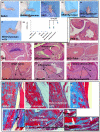
Figure 1
Figure 1
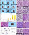
Induction of arthritis in SKG mice. (a to f) Images of the hind and fore limbs (a) control Balb/c mice (no inflammation); (b) Balb/c mice+Zymosan (no inflammation); (c) SKG mice (no inflammation); (d) SKG mice + Zymosan A (moderate inflammation); (e) SKG mice +Zymosan A + TUBO (moderate inflammation); (f) SKG mice + Zymosan A + 4T1 cells (severe inflammation). (g) Scoring of joint swelling: Compared with SKG mice + Zymosan A or SKG mice +Zymosan A + TUBO, a significant increase in joint swelling was observed in SKG mice injected with Zymosan A and challenged with 4T1 cells (P < 0.05). (h to n) H&E staining of sections from the joints: (h) Balb/c mice; (i) SKG mice; (j) SKG mice + Zymosan A without tumor challenge (moderate inflammation); (k) SKG mice + TUBO cells; (l) SKG mice + Zymosan A + TUBO cells showing some erosion of articular cartilage; (m) SKG mice + 4T1 cells showing severe inflammation in the phalangeal joints; (n) SKG mice + Zymosan A + 4T1 cells showing severe synovial hyperplasia and erosion of articular cartilage and bone in phalangeal joints with severe inflammation; (o to t) Masson trichome staining of sections from the bones: (o) Balb/c mice; (p) Balb/c mice + Zymosan A; (q) Balb/c mice +Zymosan A + 4T1; (r) SKG mice; (s) SKG mice + Zymosan A (arrow represents osteoclast); (t) SKG + Zymosan A + 4T1 (multiple osteoclasts marked with arrows). The H&E images of the joints were taken at 200 × magnification and the Masson Trichome bone images taken at 400 × magnification.
Figure 2
Figure 2
Kinetics of tumor growth in arthritic versus non-arthritic mice. (a) Mice injected with 4T1 cells; (b) mice injected with TUBO cells. Compared with the non-arthritic Balb/c mice, a significant increase in tumor burden was observed in SKG mice challenged with 4T1 (** P < 0.01) and TUBO (* P < 0.05) cells. Note: All tumor cells were injected in the mammary fat pad and sacrificed four-weeks post tumor challenge. n = 10 mice per group.
Figure 3
Figure 3
Autoimmune arthritis-related inflammation in the lungs is associated with a three-fold increase in the number of lung metastatic lesions in the SKG mice. Representative images of lungs from (a) Balb/c; (b) SKG; (c) SKG + Zymosan A; (d) Balb/c injected with TUBO cells; (e) SKG without Zymosan A + TUBO cells; (f) SKG + Zymosan A + TUBO cells (2/8 mice developed lung lesions); (g) Balb/c + 4T1 cells (3/11 mice developed lung lesions); (h) SKG without Zymosan A + 4T1 cells (8/8 mice developed severe lung metastasis); (i) SKG + Zymosan A + 4T1 cells (9/9 mice developed severe lung metastasis). (j) Percentage of mice that developed lung metastasis. (k and l) H&E of bone section showing high inflammation and image of lung with metastasis from the same 4T1-bearing SKG mouse. (m) A statistical analysis for correlation of bone inflammation and lung metastasis. The correlation coefficient for the 4T1 tumor-bearing SKG and SKG + Zymosan mice was determined to be 0.93 and 0.90. Correlation for TUBO-bearing mice was not calculated. (n to w) H&E staining of the lung sections from (n) SKG (moderate infiltration); (o) SKG + Zymosan A (moderate infiltration). (p) Balb/c + TUBO (no inflammation); (q) SKG + TUBO (moderate infiltration); (r) Balb/c + Zymosan A + TUBO; (s) SKG +Zymosan A + TUBO (moderate infiltration)(t) Balb/c + 4T1 (no infiltration); (u) SKG + 4T1 (severe infiltration); (v) Balb/c + Zymosan A + 4T1 (no infiltration); and (w) SKG + Zymosan A + 4T1 (severe infiltration). The solid filled arrow represents lung metastatic lesions, unfilled arrow represents neutrophils and unfilled circles represent macrophages. All images taken at 200 × magnification.
Figure 4
Figure 4
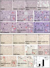
Increased infiltration of neutrophils and macrophages in the arthritic SKG mice versus the non-arthritic Balb/c mice. (a to j) Naphthol AS-D chloroacetate esterase staining of the lungs staining for neutrophils. (a) Balb/c; (b) Balb/c + Zymosan; (c) Balb/c + Zymosan + TUBO; (d) Balb/c + Zymosan + 4T1; (e) SKG (moderate infiltration of neutrophils); (f) SKG + TUBO(moderate infiltration); (g) SKG + 4T1(severe infiltration of neutrophils); (h) SKG + Zymosan(moderate infiltration); (i) SKG + Zymosan + TUBO (moderate infiltration); (j) SKG + Zymosan +4 T1(severe infiltration of neutrophils). No infiltration of neutrophils observed in any other experimental groups. (k to t) F4/80 staining of the lungs showing macrophages. (k) Balb/c; (l) Balb/c + Zymosan; (m) Balb/c + Zymosan + TUBO; (n) Balb/c + Zymosan + 4T1; (o) SKG; (p) SKG + TUBO; (q) SKG +4T1 (increased macrophages); (r) SKG + Zymosan (few macrophages); (s) SKG + Zymosan + TUBO (few macrophages); (t) SKG + Zymosan + 4T1 (increased infiltration of macrophages). No infiltrating macrophages observed in any other experimental groups. (u) BAL fluid from arthritic lungs caused significant increase in invasiveness of the 4T1 cells as compared with the BAL from non-arthritic lungs. All images taken at 200 × magnification. All slides were examined by the histology personnel and Dr Gruber at the Carolinas Medical Center confirming neutrophil infiltration.
Figure 5
Figure 5
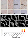
Increased expression of COX-2 and pancytokeratin in the bones of arthritic mice challenged with 4T1 cells. Bone sections from various experimental groups (indicated in the Figure) stained for COX-2 (a to f) and pancytokeratin (g to l). Brown staining represents positive staining. All images taken at 200 × magnification. (m to s) Representative Faxitron x-ray images of bones. (m) Balb/c + TUBO; (n) Balb/c + 4T1; (o) Balb/c + Zymosan + 4T1; (p) SKG + TUBO; (q) SKG + Zymosan + Tubo; (r) SKG + 4T1 (showing small radiolucencies in distal femur, proximal tibia, and head of humerus indicating apparent osteolytic bone lesions); (s) SKG + Zymosan A + 4T1 (showing radiolucencies in the distal femur, proximal tibia and head of humerus possibly due to osteolytic bone lesion). Six to seven mice from each group were examined using the faxitron x-ray. No lytic or sclerotic lesions were observed in any other experimental group. (t) Percentage of mice that developed bone metastasis.
Figure 6
Figure 6
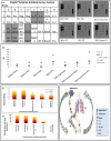
Serum analysis of cytokines revealed up regulation of several cytokines in the arthritic mice compared with the non-arthritic mice, and treatment with IL-17 antibody and celecoxib completely prevented formation of lung metastasis in the SKG mice. (a) Array template of the cytokines on the membrane. Up-regulation of macrophage colony stimulating factor (M-CSF), TNF-alpha, IL-17, vascular endothelial growth factor (VegF), and IL-6 is observed and highlighted in gray on the template (b to g) Membrane dot blot representing circulating cytokine levels in various groups of mice (indicated in the Figure). Boxes are drawn around the cytokines that were up-regulated. (h) A graphical representation of the up-regulated cytokines observed on the membrane blots based on densitometry analysis (n = 4 mice). (i) Significant reduction in tumor burden in mice treated with celecoxib (P < 0.05), α-IL-17 antibody (P < 0.01) or α-IL-17 antibody + celecoxib (P < 0.01) as compared with the untreated or IgG isotype group; (j) Percentage of mice that developed lung metastasis. (k) Schematic model depicting the vicious interaction between metastatic breast tumors and the inflammatory microenvironment in the bone and lung due to autoimmune arthritis (AA). During arthritis, inflammatory cell infiltrate in the bones and lungs trigger the release of pro-inflammatory cytokines. These cytokines act directly or indirectly on the primary breast tumor cells enhancing their metastatic ability. The ensuing damage to the bones and the large amounts of inflammatory cells in the bones and lungs of the arthritic mice allows retention and growth of the tumor cells in the site. The final distribution of metastasis reflects the relative abundance of inflammation in the organs. Thus, we hypothesize that the increase in some of the proinflammatory cytokines such as IL-17 may be the underlying factor responsible for the increased metastasis.

"VSports在线直播" References

    1. de Visser KE, Coussens LM. The inflammatory tumor microenvironment and its impact on cancer development. Contrib Microbiol. 2006;13:118–137. full_text. - PubMed
    1. Ziegler J. Cancer and arthritis share underlying processes. J Natl Cancer Inst. 1998;90:802–803. doi: 10.1093/jnci/90.11.802. - DOI - PubMed
    1. Franklin J, Lunt M, Bunn D, Symmons D, Silman A. Influence of inflammatory polyarthritis on cancer incidence and survival: results from a community-based prospective study. Arthritis Rheum. 2007;56:790–798. doi: 10.1002/art.22430. - DOI (VSports) - PubMed
    1. Mellemkjaer L, Linet MS, Gridley G, Frisch M, Moller H, Olsen JH. [Rheumatoid arthritis and risk of cancer] Ugeskr Laeger. 1998;160:3069–3073. - PubMed
    1. Askling J, Fored CM, Baecklund E, Brandt L, Backlin C, Ekbom A, Sundstrom C, Bertilsson L, Coster L, Geborek P, Jacobsson LT, Lindblad S, Lysholm J, Rantapää-Dahlqvist S, Saxne T, Klareskog L, Feltelius N. Haematopoietic malignancies in rheumatoid arthritis: lymphoma risk and characteristics after exposure to tumour necrosis factor antagonists. Ann Rheum Dis. 2005;64:1414–1420. doi: 10.1136/ard.2004.033241. - DOI - PMC - PubMed

Publication types

MeSH terms (V体育官网入口)

LinkOut - more resources (V体育ios版)