Skip to main page content
U.S. flag

An official website of the United States government

Dot gov

The . gov means it’s official. Federal government websites often end in . gov or . mil. Before sharing sensitive information, make sure you’re on a federal government site. VSports app下载.

Https

The site is secure V体育官网. The https:// ensures that you are connecting to the official website and that any information you provide is encrypted and transmitted securely. .

. 2009 Aug 1;69(15):6299-306.
doi: 10.1158/0008-5472.CAN-09-0820. Epub 2009 Jul 14.

3-Phosphoinositide-dependent kinase 1 potentiates upstream lesions on the phosphatidylinositol 3-kinase pathway in breast carcinoma

Affiliations

3-Phosphoinositide-dependent kinase 1 potentiates upstream lesions on the phosphatidylinositol 3-kinase pathway in breast carcinoma (V体育2025版)

Matthew Maurer et al. Cancer Res. .

"V体育平台登录" Abstract

Lesions of ERBB2, PTEN, and PIK3CA activate the phosphatidylinositol 3-kinase (PI3K) pathway during cancer development by increasing levels of phosphatidylinositol-3,4,5-triphosphate (PIP(3)). 3-Phosphoinositide-dependent kinase 1 (PDK1) is the first node of the PI3K signal output and is required for activation of AKT. PIP(3) recruits PDK1 and AKT to the cell membrane through interactions with their pleckstrin homology domains, allowing PDK1 to activate AKT by phosphorylating it at residue threonine-308. We show that total PDK1 protein and mRNA were overexpressed in a majority of human breast cancers and that 21% of tumors had five or more copies of the gene encoding PDK1, PDPK1. We found that increased PDPK1 copy number was associated with upstream pathway lesions (ERBB2 amplification, PTEN loss, or PIK3CA mutation), as well as patient survival. Examination of an independent set of breast cancers and tumor cell lines derived from multiple forms of human cancers also found increased PDK1 protein levels associated with such upstream pathway lesions. In human mammary cells, PDK1 enhanced the ability of upstream lesions to signal to AKT, stimulate cell growth and migration, and rendered cells more resistant to PDK1 and PI3K inhibition. After orthotopic transplantation, PDK1 overexpression was not oncogenic but dramatically enhanced the ability of ERBB2 to form tumors. Our studies argue that PDK1 overexpression and increased PDPK1 copy number are common occurrences in cancer that potentiate the oncogenic effect of upstream lesions on the PI3K pathway. Therefore, we conclude that alteration of PDK1 is a critical component of oncogenic PI3K signaling in breast cancer VSports手机版. .


Conflict of interest statement

Disclosure of Potential Conflicts of Interest

No potential conflicts of interest were disclosed.

Figures

Figure 1
Figure 1
PDK1 is overexpressed with increased genetic copy number in human breast carcinoma. (A) IHC staining for PDK1: columnar cell hyperplasia (CCH, 20x) with moderate PDK1 expression (arrow) adjacent to normal epithelium (arrowhead) reflects variable expression of PDK1 within non-neoplastic duct epithelium, Ductal carcinoma in situ (DCIS, 40x) with increased PDK1 expression (arrow), Invasive ductal carcinoma (IDC-1, 40x) with irregular cords of tumor cells over-expressing PDK1 (arrow) and adjacent normal duct (arrowhead). IDC-2 (40x) with low level of PDK1 expression (arrow) and adjacent normal duct (arrowhead). (B) Box-plot showing PDK1 IHC score distribution between BCs vs. adjacent normal ducts, as well as in tumors with PDPK1 ICN (≥5 copies) and tumors without PDPK1 ICN (≤4 copies). (C) Interphase FISH for PDPK1 (red) with centromere chromosome 16 control (green) and indicated number of PDPK1 copies. (D) Quantitative RT-PCR for PDPK1 mRNA correlated with ICN (by FISH), comparing ICN cases (relative message quantity avg.=14.0 [horizontal line]) compared to cases without ICN (avg.=8.6 [horizontal line]) among BCs with at least 50% tumor density as determined by H&E staining (n=57, Spearman coefficient of rank correlation = 0.321, p=0.016, 95% CI [0.066 to 0.537]).
Figure 2
Figure 2
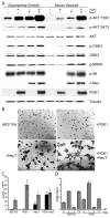
Overexpression of PDK1 enhances oncogenic phenotype in setting of upstream PI3K activation. (A) Immunoblots showing signaling effects of overexpressed PDK1 and NeuT on MCF10A cells under normal exponential growth or growth factor withdrawal conditions. (B) Matrigel morphogenesis assay of stably transfected pools of MCF10A cells as indicated. (C) Transwell migration assay with same set of cells with and without chemo attractant (epidermal growth factor and 5% horse serum) (s.d., n=3). (D) Migration assay of control MCF10A cells or those with PDK1 overexpression with siRNA knockdown of AKT1, AKT2, or both. Migration (y-axis) = ratio of the number of migratory cells in test vs. control (ctrl) (s.d., n=3).
Figure 3
Figure 3
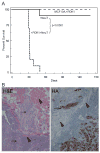
Increased PDK1 potentiates tumor growth in vivo. (A) Kaplan-Meier survival curves of MCF10A cells injected into the mammary fatpads of developing scid mice comparing cells overexpressing NeuT with (+PDK1+NeuT, narrow dashed line) or without (+NeuT, solid line) overexpressed PDK1 [death is defined as tumor growth to size = 1 cm2]. Injected control MCF10A cells and cells overexpressing PDK1 alone did not form tumors (wide dashed line). (B) IHC staining (left panel=H&E, right panel=HA antibody to HA tagged PDK1, 40x) of xenografted tumor cells (arrows) invading host muscle (arrowheads).
Figure 4
Figure 4
In cells with PI3K activation, PDK1 levels are a determinant of signaling, proliferation, transformation, and pathway inhibition. (A) Immunoblots of MCF7 cells grown with stable control shRNA [CTRL] or two separate PDK1 shRNA contructs [PDK1-1 and PDK1-2]. (B) MCF7 cells with stable control shRNA or separate PDK1 shRNA contructs (as indicated) grown for four days with 1.25 μM BX-795 (orange bars) or control (DMSO, blue bars) (percent inhibition indicated, s.d., n=3). (C) BX-795 dose response curves of control MCF7 cells or MCF7 cells over-expressing PDK1 with the IC50 indicated (dotted lines) (s.d., n=3). (D) Colony formation assay of HMEC/hTERT/p53DD cells over-expressing mutant p110α (H1047R) or control, or p110α (H1047R) in the setting of stable PDK1 shRNA compared with control shRNA.

References

    1. Isakoff SJ, Engelman JA, Irie HY, et al. Breast cancer-associated PIK3CA mutations are oncogenic in mammary epithelial cells. Cancer Res. 2005;65:10992–1000. - "VSports注册入口" PubMed
    1. Li J, Simpson L, Takahashi M, et al. The PTEN/MMAC1 tumor suppressor induces cell death that is rescued by the AKT/protein kinase B oncogene. Cancer Res. 1998;58:5667–72. - "VSports最新版本" PubMed
    1. Stambolic V, Suzuki A, de la Pompa JL, et al. Negative regulation of PKB/Akt-dependent cell survival by the tumor suppressor PTEN. Cell. 1998;95:29–39. - PubMed
    1. Zhao JJ, Liu Z, Wang L, Shin E, Loda MF, Roberts TM. The oncogenic properties of mutant p110alpha and p110beta phosphatidylinositol 3-kinases in human mammary epithelial cells. Proc Natl Acad Sci U S A. 2005;102:18443–8. - PMC - PubMed
    1. Zhou BP, Hu MC, Miller SA, et al. HER-2/neu blocks tumor necrosis factor-induced apoptosis via the Akt/NF-kappaB pathway. J Biol Chem. 2000;275:8027–31. - PubMed

"VSports最新版本" Publication types

MeSH terms

"V体育平台登录" Substances

LinkOut - more resources