Skip to main page content
U.S. flag

An official website of the United States government

Dot gov

The . gov means it’s official. Federal government websites often end in VSports app下载. gov or . mil. Before sharing sensitive information, make sure you’re on a federal government site. .

Https

The site is secure. The https:// ensures that you are connecting to the official website and that any information you provide is encrypted and transmitted securely. V体育官网.

. 2009 Jul;8(7):3497-511.
doi: 10.1021/pr9001614.

Proteomic studies of nitrated alpha-synuclein microglia regulation by CD4+CD25+ T cells

Affiliations

Proteomic studies of nitrated alpha-synuclein microglia regulation by CD4+CD25+ T cells

Ashley D Reynolds et al. J Proteome Res. 2009 Jul.

Abstract

Microglial inflammatory responses affect Parkinson's disease (PD) associated nigrostriatal degeneration VSports手机版. This is triggered, in measure, by misfolded, nitrated alpha-synuclein (N-alpha-syn) contained within Lewy bodies that are released from dying or dead dopaminergic neurons into the extravascular space. N-alpha-syn-stimulated microglial immunity is regulated by CD4+ T cell subset. Indeed, CD4+CD25+ regulatory T cells (Treg) induce neuroprotective immune responses. This is seen in rodent models of stroke, amyotrophic lateral sclerosis, human immunodeficiency virus associated neurocognitive disorders, and PD. To elucidate the mechanism for Treg-mediated microglial neuroregulatory responses, we used a proteomic platform integrating difference gel electrophoresis and tandem mass spectrometry peptide sequencing. These tests served to determine consequences of Treg on the N-alpha-syn stimulated microglia. The data demonstrated that Treg substantially alter the microglial proteome in response to N-alpha-syn. This is seen through Treg abilities to suppress microglial proteins linked to cell metabolism, migration, protein transport and degradation, redox biology, cytoskeletal, and bioenergetic activities. We conclude that Treg modulate the N-alpha-syn microglial proteome and, in this way, can slow the tempo and course of PD. .


Figures
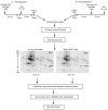
Figure 1
Figure 1
Experimental design for microglial proteomics protein discovery. Microglia were co-cultured for 24 h with CD4+CD25+ Treg (or Teff) or without as control. Treg (or Teff) were removed from the cultures and the microglia stimulated with aggregated N-α-syn for 24 h [pre-treatment] to represent asymptomatic disease (A). Alternatively, microglia were stimulated with N-α-syn for 12 h prior to the addition of Treg [post-treatment] to represent more overt disease (B). Twenty-four hours later microglial cell protein lysates were prepared and subjected to 2D electrophoresis. Decyder analysis software was used to match spots and identify expression patterns. Selected protein spots were excised, digested with trypsin and identified by nano-LC-MS/MS peptide sequencing. Database searches were performed using SEQUEST with criteria thresholds set to afford greater than 95% confidence level in peptide identification.
Figure 2
Figure 2
2D-DIGE of proteins from all experimental groups with matched spots picked for sequencing identifications by nano-LC-MS/MS. A representative preparative gel is shown. Equal amounts of protein were pooled from all experimental groups (unstimulated, N-α-syn-stimulated, pre- and post-treatment with Treg or Teff) and replicates for a total concentration of 450 μg. The pooled sample was applied to a pH gradient strip and separated with isoelectric focusing for the first dimension. For the second dimension, the strip was loaded onto a large format gradient gel and separated based on molecular weight. Following electrophoresis, the gel was fixed and post-stained with Deep Purple for positive detection of protein spots. Circled spots identified by BVA using Decyder analysis software were selected for excision. Proteins with the most peptides positively identified within a specific spot are labeled on the gel. (Abbreviations: Prdx, peroxiredoxin; Thx, thioredoxin, Vdac, voltage-dependent anion channel; Sod, superoxide dismutase; Hsp, heat shock protein; Capg, macrophage capping protein; NAD, nicotinamide adenine dinucleotide).
Figure 3
Figure 3
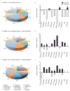
Classification of proteins modulated by N-α-syn stimulation and Treg treatment. Pie-chart diagrams represent the proportion (%) of proteins within specific categories based on classification and function identified by mass spectrometry. (A) Classification of proteins differentially expressed by microglia in response to N-α-syn stimulation alone. (B) Relative expression of proteins in response to N-α-syn stimulation compared to unstimulated controls. Several proteins within each category showing both increased and decreased proteins were identified including those for apoptosis (*gelsolin increased; nucleoside-diphosphate kinase decreased) and glycolysis (§enolase 3 and lactate dehydrogenase increased; alpha enolase, pyruvated dehydrogenase, pyruvate kinase, and triosphosphate isomerase 1 decreased) (Table 1). (C) Proportion of microglial proteins differentially expressed in response to N-α-syn following Treg pre-treatment and the relative expression trends shown in D. Categories associated with transcription (*VIP-receptor gene repressor protein, TAR DNA binding protein, and Ubiquitin conjugating enzyme E2N increased; MRG-binding protein decreased), cell motility (§microtubule associated protein increased; laminin B2, beta actin, and alpha tubulin decreased), structural (#Capg and guanine nucleotide exchange factor increased; vimentin, cofilin 1 and 2 decreased), and oxidative phosphorylation (NADH dehydrogenase Fe-S, ATP synthase O subunit, H+-ATP synthase e subunit, and cytochrome c oxidase increased; ATP synthase F0 complex decreased) consisted of both increased and decreased expression of proteins (Table 2). (E) Proportion of microglial proteins differentially expressed in response to N-α-syn following Treg post-treatment and the relative expression trends shown in F. Categories associated with metabolism (#phosphoglycerate mutase 1 increased; aldolase 1 and aldehyde dehydrogenase 2 decreased), oxidative phosphorylation (§ATP synthase D increased; H+-transporting two-sector ATPase alpha chain decreased), and chaperones (#cyclophilin A increased; protein disulfide isomerase decreased) consisted of both increased and decreased expression of proteins (Table 3). (Proteins within this category were not identified as differentially expressed).
Figure 4
Figure 4
Treg modulate microglial inflammation to attenuate the neurotoxic phenotype of N-α-syn stimulated microglia. (A) Photomicrographs (20× magnification) of Prx1 expression (green) in microglia treated with media alone (CON), N-α-syn, or cultured with CD4+ T cell subsets following pre-and post-treatment. Values shown are the mean fluorescence intensity (MFI) per field ± SEM. (B) Western blot analysis for α-tubulin, galectin 3 and gelsolin in response to treatment normalized to Gapdh expression within the same blot for comparisons. (C) Photomicrographs (20× magnification) of actin expression (red) or Hsp70 (green) in microglia treated with media alone (CON), N-α-syn, or cultured with CD4+ T cell subsets following pre-and post-treatment. Values shown are the MFI per field ± SEM. (D) Survival of MES23.5 cells after co-culture with N-α-syn stimulated microglia with and without Treg or Teff or after culture with condition media (supernatants) of N-α-syn stimulated microglia treated with either Treg or Teff. Values ± SEM (P< 0.01 vs. aCON, bN-α-syn alone, cN-α-syn/Teff).

References

    1. Dauer W, Przedborski S. Parkinson's disease: mechanisms and models. Neuron. 2003;39(6):889–909. - "V体育官网" PubMed
    1. Fahn S, Przedborski S. Parkinsonism. In: Rowland LP, editor. Merritt's Neurology. Lippincott Williams & Wilkins; New York: 2000. pp. 679–693.
    1. Fahn S, Sulzer D. Neurodegeneration and neuroprotection in Parkinson disease. NeuroRx. 2004;1(1):139–54. - PMC - PubMed
    1. Mayeux R. Epidemiology of neurodegeneration. Annu Rev Neurosci. 2003;26:81–104. - PubMed
    1. Baba Y, Kuroiwa A, Uitti RJ, Wszolek ZK, Yamada T. Alterations of T-lymphocyte populations in Parkinson disease. Parkinsonism Relat Disord. 2005;11(8):493–8. - "VSports在线直播" PubMed

Publication types

MeSH terms