Skip to main page content
U.S. flag

An official website of the United States government

Dot gov

The . gov means it’s official. Federal government websites often end in . gov or VSports app下载. mil. Before sharing sensitive information, make sure you’re on a federal government site. .

Https

The site is secure. The https:// ensures that you are connecting to the official website and that any information you provide is encrypted and transmitted securely. V体育官网.

. 2009 May;58(5):1050-7.
doi: 10.2337/db08-1299. Epub 2009 Feb 2.

MicroRNAs induced during adipogenesis that accelerate fat cell development are downregulated in obesity

Affiliations

MicroRNAs induced during adipogenesis that accelerate fat cell development are downregulated in obesity

Huangming Xie et al. Diabetes. 2009 May.

Abstract

Objective: We investigated the regulation and involvement of microRNAs (miRNAs) in fat cell development and obesity VSports手机版. .

Research design and methods: Using miRNA microarrays, we profiled the expression of >370 miRNAs during adipogenesis of preadipocyte 3T3-L1 cells and adipocytes from leptin deficient ob/ob and diet-induced obese mice. Changes in key miRNAs were validated by RT-PCR. We further assessed the contribution of the chronic inflammatory environment in obese adipose tissue to the dysregulated miRNA expression by tumor necrosis factor (TNF)-alpha treatment of adipocytes. We functionally characterized two adipocyte-enriched miRNAs, miR-103 and miR-143, by a gain-of-function approach. V体育安卓版.

Results: Similar miRNAs were differentially regulated during in vitro and in vivo adipogenesis V体育ios版. Importantly, miRNAs that were induced during adipogenesis were downregulated in adipocytes from both types of obese mice and vice versa. These changes are likely associated with the chronic inflammatory environment, since they were mimicked by TNF-alpha treatment of differentiated adipocytes. Ectopic expression of miR-103 or miR-143 in preadipocytes accelerated adipogenesis, as measured both by the upregulation of many adipogenesis markers and by an increase in triglyceride accumulation at an early stage of adipogenesis. .

Conclusions: Our results provide the first experimental evidence for miR-103 function in adipose biology. The remarkable inverse regulatory pattern for many miRNAs during adipogenesis and obesity has important implications for understanding adipose tissue dysfunction in obese mice and humans and the link between chronic inflammation and obesity with insulin resistance VSports最新版本. .


Figures
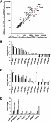
FIG. 1.
FIG. 1.
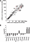
miRNA expression profiling during 3T3-L1 adipogenesis. A: Intensity scatter plot showing comparison of miRNA profiles between undifferentiated 3T3-L1 preadipocytes (day 0) and differentiated 3T3-L1 adipocytes (day 9). The 12 miRNAs that are the focus of our detailed analyses are highlighted in red and indicated by a label. B: Validation of miRNA array results for 12 regulated miRNAs by RT-PCR assays. Expressions for all miRNAs are plotted as fold-change in log2 scale; positive indicates enriched in day 9 vs. day 0. □, Array result (n = 2); ■, RT-PCR result (n = 4). Data are expressed as means ± SE. C: Comparison by RT-PCR of miRNA regulation during 3T3-L1 differentiation and primary fat cell development. Expressions of all miRNAs are normalized to internal control and plotted as fold-change in log2 scale. ■, day 9 vs. day 0, same data as Fig. 1B (n = 4); □, mature adipocytes vs. enriched preadipocytes (n = 4). Data are expressed as mean ± SE. D: Intronic miRNAs and their host genes are co-regulated during adipogenesis. miR-422b is in the intron of PPARGC1b; miR-107 is in the intron of PANK1; the two copies of miR-103 are in the introns of PANK2 and PANK3. miRNA expression levels are measured by RT-PCR and normalized to internal control. Expression levels of mRNAs are also determined by RT-PCR and normalized to internal control. Expressions are shown as fold-change relative to the level at day 0. □, day 0; ■, day 2; ▨, day 4. Data are expressed as mean ± SE (n = 3).
FIG. 2.
FIG. 2.
miRNA expression profiling in adipocytes from wild-type (WT) and leptin-deficient ob/ob mice. A: Intensity scatter plot showing comparison of miRNA profiles in adipocytes from WT and ob/ob mice. The 12 miRNAs that are the focus of our detailed analyses are highlighted in red and indicated by a label. B: Validation of miRNA array results for 12 regulated miRNAs by RT-PCR assays. Expression of all miRNAs are normalized to internal control and plotted as fold-change in a log2 scale. □, Array result (n = 2); ■, RT-PCR result (n = 2). Data are expressed as mean ± SE.
FIG. 3.
FIG. 3.
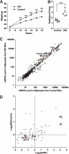
miRNA expression profiling in adipocytes from control and DIO mice. A: Weight of control and DIO mice (n = 5). DIO mice were fed with a 55% high-fat diet starting from week 6 to week 18. Controls were fed with a normal diet. Data are expressed as means ± SE. **P < 0.01. B: Weight of epididymal fat pads from control and DIO mice (n = 5) at week 18. **P < 0.01. C: Intensity scatter plot showing comparison of miRNA profiles in adipocytes from control and DIO mice. Mirtron miR-1224, Novel_miR#5 (novel miRNA cloned from bovine adipose tissue described in the text), and some miRNAs used in our detailed analyses are highlighted in red and indicated by a label. D: Scatter plot showing the positive correlation of miRNA regulation in two disease models for obesity: ob/ob and DIO mice. A total of 78 miRNAs, whose levels are above the mean expression of all miRNAs in at least one of the four samples (enriched adipocytes from wild-type [WT], ob/ob, control, and DIO mice), are shown. Fold-changes are based on array results and plotted in a log2 scale. Some miRNAs used in our detailed analyses are highlighted in red and indicated by a label. Pearson's correlation coefficient: r = 0.51, P < 0.0001.
FIG. 4.
FIG. 4.
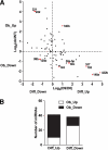
Inverse correlation of miRNA expression during adipogenesis and obesity. A: Scatter plot showing the inverse correlation of miRNA regulation during adipogenesis and obesity. A total of 79 miRNAs, whose levels are above the mean expression of all miRNAs in at least one of the four samples (undifferentiated 3T3-L1 preadipocytes [day 0], differentiated 3T3-L1 adipocytes [day 9], and adipocytes from wild-type [WT] and ob/ob mice) are shown. Fold-change is based on array result and plotted in a log2 scale. The 12 miRNAs that are the focus of our detailed analyses are highlighted in red and indicated by a label. Pearson's correlation coefficient: r = −0.51, P < 0.0001. B: Fisher's exact test for 79 miRNAs is shown in Fig. 3A. A total of 41 miRNAs were upregulated during differentiation (Diff_Up), among which 31 miRNAs were downregulated during obesity (Ob_Down), whereas 10 miRNAs were upregulated (Ob_Up). A total of 38 miRNAs were downregulated during differentiation (Diff_Down), among which 26 miRNAs were upregulated during obesity (Ob_Up), whereas 12 miRNAs were downregulated (Ob_Down). P = 0.0001 by Fisher's exact test.
FIG. 5.
FIG. 5.
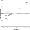
miRNAs differentially expressed in adipocytes from ob/ob mice compared with wild-type (WT) mice are regulated similarly by TNF-α treatment of 3T3-L1 differentiated adipocytes for 24 h. miRNA levels are measured by RT-PCR, normalized to internal control and plotted as fold-change in log2 scale. x-axis, ob/ob vs. WT, same data as Fig. 2B; y-axis, differentiated adipocyte after TNF-α treatment for 24 h versus untreated control (n = 4). Pearson's correlation coefficient: r = 0.90, P < 0.0001.
FIG. 6.
FIG. 6.
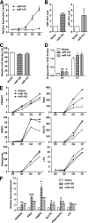
Ectopic expression of two adipocyte-induced miRNAs, miR-103 or miR-143, accelerates adipogenesis. A: miR-103 and miR-143 are induced during 3T3-L1 adipogenesis. miRNA expression levels are measured by RT-PCR, normalized to internal control and plotted relative to their respective levels in preadipocyte (day 0). Data are expressed as mean ± SE (n = 3). B: Expression level of miR-103 or miR-143 in infected and sorted cells compared with vector control. Data are expressed as mean ± SE (n = 3). C: Ectopic expression of miR-103 or miR-143 has little effect on 3T3-L1 cell growth by MTT assay. Data are expressed as mean ± SE (n = 6). D: Ectopic expression of miR-103 or miR-143 increases triglyceride accumulation at day 4 (D4) but not day 7 (D7). Triglycerides are normalized to total protein and plotted relative to the level of the control cells expressing vector alone at D7. Data are expressed as mean ± SE (n = 6). **P < 0.01, *P < 0.05. E: Ectopic expression of miR-103 or miR-143 hastens expression of adipogenic markers at an early stage (days 2 and 4) of adipogenesis. mRNA levels are measured by RT-PCR, normalized to internal control and plotted relative to their respective levels at day 2 (D2). Data are expressed as mean only (n = 3). PPARG2, peroxisome proliferator-activated receptor γ 2; G0s2, G0/G1 switch 2; FABP4, fatty acid binding protein 4; GLUT4, glucose transporter 4; LPL, lipoprotein lipase. F: Ectopic expression of miR-103 or miR-143 hastens expression of adipogenic markers at day 2 (D2). Data are expressed as mean ± SE (n = 4). ***P < 0.001, **P < 0.01, *P < 0.05 by one-sample Student's t test.

References

    1. Rosen ED, Spiegelman BM: Adipocytes as regulators of energy balance and glucose homeostasis. Nature 2006; 444: 847– 853 - PMC - PubMed
    1. Rajala MW, Scherer PE: Minireview: the adipocyte: at the crossroads of energy homeostasis, inflammation, and atherosclerosis. Endocrinology 2003; 144: 3765– 3773 - PubMed
    1. Schaffler A, Muller-Ladner U, Scholmerich J, Buchler C: Role of adipose tissue as an inflammatory organ in human diseases. Endocr Rev 2006; 27: 449– 467 - PubMed
    1. Calle EE, Kaaks R: Overweight, obesity and cancer: epidemiological evidence and proposed mechanisms. Nat Rev Cancer 2004; 4: 579– 591 - PubMed
    1. Kopelman PG: Obesity as a medical problem. Nature 2000; 404: 635– 643 - PubMed

Publication types

MeSH terms