<tt lang="KMm35lMy"></tt> Skip to main page content
U.S. flag

An official website of the United States government

Dot gov

The . gov means it’s official. Federal government websites often end in . gov or . mil. Before sharing sensitive information, make sure you’re on a federal government site VSports app下载. .

Https

The site is secure. The https:// ensures that you are connecting to the official website and that any information you provide is encrypted and transmitted securely. V体育官网.

Comparative Study
. 2009 Jan 1;182(1):148-53.
doi: 10.4049/jimmunol.182.1.148.

Levels of Foxp3 in regulatory T cells reflect their functional status in transplantation

Affiliations
Comparative Study

"V体育官网" Levels of Foxp3 in regulatory T cells reflect their functional status in transplantation

Sunil K Chauhan (VSports在线直播) et al. J Immunol. .

Abstract

Foxp3 expressing CD4(+)CD25(+) regulatory T cells (Tregs) have been shown to prevent allograft rejection in clinical and animal models of transplantation. However, the role of Foxp3 in regulating Treg function, and the kinetics and mechanism of action of Tregs in inducing allograft tolerance in transplantation, are still not fully understood. Thus, we investigated the kinetics and function of Tregs in a mouse model of orthotopic corneal transplantation, the most common form of tissue grafting worldwide. In this study, using in vitro functional assays and in vivo Treg adoptive transfer assays, we show that far more relevant than Treg frequency is their level of Foxp3 expression, which is directly associated with the potential of Tregs to prevent allograft rejection by producing regulatory cytokines and suppressing effector T cell activation VSports手机版. In addition, our data clearly demonstrate that Tregs primarily suppress the induction of alloimmunity in regional draining lymph nodes rather than suppressing the effector phase of the immune response in the periphery. These findings provide new insights on Treg dynamics in transplantation, which are crucial for designing therapeutic strategies to modulate Treg function and to optimize Treg-based cell therapies for clinical translation. .


Figures
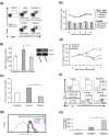
FIGURE 1
FIGURE 1. Kinetics of Treg frequency and Foxp3 expression in syngeneically grafted recipients (Balb/c→Balb/c) and fully disparate allograft (C57Bl/6→Balb/c) acceptors and rejectors
(a) FACS analysis of draining lymph nodes (LN) cell suspension showing frequencies of CD4+CD25+Foxp3+ Tregs at week-3 post transplantation. (b) Frequencies of Tregs out of CD4+ and total LN cells at weekly intervals from Day 0 to week 4 post-transplantation. (c) Mean fluorescence intensity and western blot analyses showing expression levels of Foxp3 protein in purified Tregs isolated from the LN at wk-3 post-transplantation (*p = 0.036). (d) Real-time PCR analysis of Foxp3 mRNA expression levels in total LN cells at weekly intervals from Day 0 to week 4 post-transplantation (acceptor vs. syngeneic and rejector at wk-3 *p = 0.034 and 0.027; and at wk-4 p = 0.01 and 0.001); and (e) in equivalent numbers of purified Tregs sorted from the LN at week-3 post transplantation (*p = 0.026). (f) Frequencies of corneal graft-infiltrating Foxp3+ Tregs and total CD3+ T cells and (g) MFI of Foxp3 levels in graft-infiltrating Tregs. (h) Real-time PCR analysis of Foxp3 mRNA expression in the grafted corneas of syngeneically graft recipients, allograft acceptors and allograft rejectors at week 3 post-transplantation (*p = 0.037). Each transplant group consists of 4-6 recipient mice and data present the mean ± SEM of three independent experiments. Statistical significance for mRNA and MFI levels is calculated on fold-change values over those of syngeneic or naïve-Tregs. P values are calculated using student’s t-test.
FIGURE 2
FIGURE 2. Suppressor potential of Tregs
(a) Naïve CD4+T cells isolated from the LN of unprimed BALB/c recipients and (b) allo-primed CD4+T cells isolated from the LN of allograft rejectors were stimulated with CD3-antibody for 3 days in the presence of different Tregs isolated from the LN of syngeneically grafted recipients, allograft acceptors, and allograft rejectors at week 3 post-transplantation. The activity of Tregs is measured at Treg:Teff cell ratio of 1:2. Proliferation was measured using the BrdU incorporation assay and compared with the proliferative responses of respective CD3-stimulated naïve or allo-primed T cell in the absence of Tregs and % suppression was calculated as described in Materials and Methods. Each transplant group consists of 4-6 recipient mice. Data from a representative experiment of three performed is shown. P values are calculated using student’s t-test and error bars represent SEM. *p = 0.018; §p = 0.0002.
FIGURE 3
FIGURE 3. Mechanism(s) of suppression utilized by Tregs
(a) ELISA based analysis of cytokines in the supernatant collected at Day 3 from co-cultures of CD3 stimulated naïve T cells isolated from the LN of unprimed BALB/c recipients with the LN-Tregs of different transplant groups (TGF-β1: *p = 0.046, §p = 0.011; IL-10: *p = 0.003, §p = 0.017). (b) In transwell assays, naïve T cells isolated from the LN of unprimed BALB/c recipients were stimulated with anti-CD3 antibody, and the LN-Tregs from different groups were either added directly to the culture or were placed in transwell inserts of 1.0μm pore size (*p = 0.01). Proliferation was measured using the BrdU incorporation assay and compared with the proliferative responses of CD3-stimulated naïve T cell in the absence of Tregs and % suppression was calculated as described in Materials and Methods. Tregs were isolated from the draining lymph nodes of syngeneic recipients, allograft acceptors, and allograft rejectors at week 3 post-transplantation. The activity of Tregs is measured at Treg:Teff cell ratio of 1:2. Each transplant group consists of 4-6 recipient mice. Data from a representative experiment of three performed is shown. P values are calculated using student’s t-test and error bars represent SEM.
FIGURE 4
FIGURE 4. Alloantigen-specificity of Tregs
The suppression potential of regulatory T cells was evaluated against proliferation of recipient’s naïve T cells to donor-specific and third-party (allodisparate to both recipient and donor) antigens. Naïve T cells from the LN of unprimed BALB/c mice (graft recipients) were co-cultured with antigen presenting cells (APC) of C57BL6 mice (graft donors) or C3H mice (third-party) in the presence of Tregs isolated form the draining lymph nodes of syngeneic recipients, allograft acceptors, and allograft rejectors at week 3 post-transplantation (*p = 0.012, Acceptor-Tregs: C57BL6-APC vs. C3H-APC). Proliferation was measured using the BrdU incorporation assay and compared with the proliferative responses of respective APC stimulated naïve T cell in the absence of Tregs and % suppression was calculated as described in Materials and Methods. Each transplant group consists of 4-6 recipient mice. Data from a representative experiment of three performed is shown. P values are calculated using student’s t-test and error bars represent SEM.
FIGURE 5
FIGURE 5. Effect of Treg adoptive transfer on allograft survival
Tregs were isolated from the LN of syngeneically grafted recipients (syngeneic Treg), allograft acceptors (acceptor Treg) and allograft rejectors (rejector Treg) at week 3 post-transplantation; 1×10⁵ Tregs per recipients were intravenously transferred to three different groups of allograft (C57BL6→ BALB/c) recipients at 18h post-surgery. Graft survival was monitored up to 8-wk post-transplantation. Controls recipients were syngeneically grafted (BALB/c→BALB/c) and received no Tregs. (n = 6 per group; *p = 0.035, Kaplan-Meier survival analysis).

References

    1. Wood KJ, Sakaguchi S. Regulatory T cells in transplantation tolerance. Nat. Rev. Immunol. 2003;3:199–210. - PubMed
    1. Kang SM, Tang Q, Bluestone JA. CD4+CD25+ regulatory T cells in transplantation: progress, challenges and prospects. Am. J. Transplant. 2007;7:1457–1463. - PubMed
    1. Sakaguchi S. Naturally arising Foxp3-expressing CD25+CD4+ regulatory T cells in immunological tolerance to self and non-self. Nat. Immunol. 2005;6:345–352. - PubMed (V体育平台登录)
    1. Hori S, Nomura T, Sakaguchi S. Control of regulatory T cell development by the transcription factor Foxp3. Science. 2003;299:1057–1061. - PubMed
    1. Joffre O, Santolaria T, Calise D, Al Saati T, Hudrisier D, Romagnoli P, van Meerwijk JP. Prevention of acute and chronic allograft rejection with CD4+CD25+Foxp3+ regulatory T lymphocytes. Nat. Med. 2008;14:88–92. - PMC - PubMed

Publication types (VSports在线直播)

MeSH terms