"VSports最新版本" Skip to main page content
U.S. flag

An official website of the United States government

Dot gov

The . gov means it’s official. Federal government websites often end in . gov or VSports app下载. mil. Before sharing sensitive information, make sure you’re on a federal government site. .

Https

The site is secure. The https:// ensures that you are connecting to the official website and that any information you provide is encrypted and transmitted securely. V体育官网.

. 2009 Feb 6;284(6):3823-32.
doi: 10.1074/jbc.M807869200. Epub 2008 Dec 15.

"V体育ios版" Role of sirtuin histone deacetylase SIRT1 in prostate cancer. A target for prostate cancer management via its inhibition?

Affiliations

Role of sirtuin histone deacetylase SIRT1 in prostate cancer. A target for prostate cancer management via its inhibition?

Brittney Jung-Hynes et al. J Biol Chem. .

Abstract

Prostate cancer (PCa) is a major age-related malignancy, and according to estimates from the American Cancer Society, a man's chance of developing this cancer significantly increases with increasing age, from 1 in 10,149 by age 39 to 1 in 38 by age 59 to 1 in 7 by age 70. Therefore, it is important to identify the causal connection between mechanisms of aging and PCa. Employing in vitro and in vivo approaches, in this study, we tested the hypothesis that SIRT1, which belongs to the Sir2 (silent information regulator 2) family of sirtuin class III histone deacetylases, is overexpressed in PCa, and its inhibition will have antiproliferative effects in human PCa cells. Our data demonstrated that SIRT1 was significantly overexpressed in human PCa cells (DU145, LNCaP, 22Rnu1, and PC3) compared with normal prostate epithelial cells (PrEC) at protein, mRNA, and enzymatic activity levels. SIRT1 was also found to be overexpressed in human PCa tissues compared with adjacent normal prostate tissue VSports手机版. Interestingly, our data demonstrated that SIRT1 inhibition via nicotinamide and sirtinol (at the activity level) as well as via short hairpin RNA-mediated RNA interference (at the genetic level) resulted in a significant inhibition in the growth and viability of human PCa cells while having no effect on normal prostate epithelial cells. Further, we found that inhibition of SIRT1 caused an increase in FOXO1 acetylation and transcriptional activation in PCa cells. Our data suggested that SIRT1, via inhibiting FOXO1 activation, could contribute to the development of PCa. We suggest that SIRT1 could serve as a target toward developing novel strategies for PCa management. .


Figures
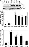
FIGURE 1.
FIGURE 1.
SIRT1 protein and mRNA levels in PCa cell lines versus normal prostate epithelial cells. A, western blot analysis of SIRT1. The cells were grown to 80% confluence, and cell lysates were prepared. SIRT1 protein levels were determined by Western blot analysis. Equal loading was confirmed by reprobing the blot for β-actin. B, quantitation of SIRT1 protein levels. Western blot analysis was quantitated by densitometric analysis of protein bands. The data (relative density normalized to β-actin) are expressed as mean ± S.E. of three experiments. *, p < 0.01. C, SIRT1 mRNA. The relative expression of SIRT1 transcript in PCa cell lines was determined by quantitative real time reverse transcription PCR using ABI-PRISM SDS software and comparative Ct methods analysis. The data is expressed as mean ± S.E. of three experiments. *, p < 0.01. Details of the experiments are given under “Experimental Procedures.”
FIGURE 2.
FIGURE 2.
SIRT1 activity in PCa cell lines versus normal prostate epithelial cells. SIRT1 protein was immunoprecipitated from protein lysate (500 μg of protein), and SIRT1 enzyme activity was assessed using the SIRT1 activity assay kit (Biomol) as per the vendor's protocol. Pure SIRT1 enzyme (SIRT1) and nicotinamide (Nic) were used as a positive and negative control, respectively. SIRT1 activity is represented as arbitrary fluorescence units (AFU). The data are expressed as mean ± S.E. of three experiments. *, p < 0.01.
FIGURE 3.
FIGURE 3.
Immunohistochemical analysis of SIRT1 in prostate tissues from patients with PCa. Immunohistochemical analysis for SIRT1 was performed on a custom made tissue microarray containing normal or benign prostate epithelium, high grade prostatic intraepithelial neoplasia, and prostate cancer samples with different Gleason patterns as well as on paraffin embedded tissue slides containing human prostate cancer tissue with adjacent normal prostate tissues. Details of the experiment are given under “Experimental Procedures.”
FIGURE 4.
FIGURE 4.
Effect of nicotinamide treatment on growth and viability of human PCa cells and normal prostate epithelial cells. A, effect of nicotinamide on SIRT1 activity. PCa cells (60% confluent) were treated with 150 μm nicotinamide or untreated control for 24 h. SIRT1 protein was immunoprecipitated using a SIRT1 antibody. SIRT1 enzyme activity was assessed using SIRT1 activity assay kit and is represented as arbitrary fluorescence units (AFU) as previously stated. The data are expressed as mean ± S.E. of three experiments. *, p < 0.01. B, effect of nicotinamide on cell growth and cell viability in normal prostate epithelial cells. The normal human prostate epithelial cells were treated with nicotinamide (150 or 300 μm nicotinamide) for 24 h and analyzed by a trypan blue assay to assess cell growth and viability. Cell growth is expressed as percentage of growth (from total number of cells), and cell viability is expressed as the percentage of viable cells of the total number of cells. The data are expressed as the mean ± S.E. of three experiments. C, effect of nicotinamide on cell growth in PCa cells. PCa cells were treated with nicotinamide (150 μm, 300 μm, 5 mm, or 20 mm nicotinamide) for 24 h and analyzed by a trypan blue assay to assess cell growth, which is expressed as percentage of cell growth (from total number of cells). The data are expressed as the mean ± S.E. of three experiments. *, p < 0.01. D, effect of nicotinamide on cell viability in PCa cells. PCa cells were treated and analyzed using a trypan blue assay as described above. Cell viability is expressed as the percentage of viable cells of the total number of cells. The data represents mean ± S.E. of three experiments. *, p < 0.01. Details of the experiments are given under “Experimental Procedures.”
FIGURE 5.
FIGURE 5.
Effect of sirtinol treatment on growth and viability of human PCa cells. A, effect of sirtinol on cell growth in PCa cells. PCa cells were treated with 30 μm or 120 μm sirtinol (dissolved in DMSO) for 24 or 48 h and analyzed by trypan blue assay to assess cell growth, which is expressed as percentage of cell growth (from total number of control cells). The data are expressed as the mean ± S.E. of three experiments. *, p < 0.01. B, effect of sirtinol on cell viability in PCa cells. PCa cells were treated with 30 or 120 μm sirtinol for 24 or 48 h and analyzed using the trypan blue assay as described above. Cell viability is expressed as the percentage of viable cells of the total number of control (DMSO-treated) cells. The data represent mean ± S.E. of three experiments. *, p < 0.01.
FIGURE 6.
FIGURE 6.
Effect of shRNA-mediated knockdown of SIRT1 on growth and viability of human PCa cells. A, effect of shRNAs on SIRT1 protein levels. Following transfection of PCa cells with SIRT1 shRNA or control nonsense shRNA (for 48 h), SIRT1 protein levels were detected by Western blot analysis. Equal loading was confirmed by reprobing the blot for β-actin. Data represent three experiments with similar results. B, quantitation of SIRT1 protein levels. Western blot data were quantitated by a densitometric analysis of protein bands. The data (relative density normalized to β-actin) are expressed as mean ± S.E. of three experiments. *, p < 0.01. C, effect of SIRT1 knockdown on SIRT1 activity. PCa cells were transfected with SIRT1 shRNA or control shRNA (for 48 h). SIRT1 protein was immunoprecipitated using a SIRT1 antibody. SIRT1 enzyme activity was assessed using a SIRT1 activity assay kit (AK-555; Biomol). The data (relative-fold change in arbitrary fluorescence units normalized to nonsense control) are expressed as mean ± S.E. of three experiments. *, p < 0.01. D, effect of SIRT1 knockdown on cell growth. The effect of SIRT1 knockdown on cell growth was analyzed by a trypan blue assay and is expressed as percentage of growth (from total number of cells). The data are expressed as the mean ± S.E. of three experiments. *, p < 0.01. E, effect of SIRT1 knockdown on cell viability. The effect of SIRT1 knockdown was measured using a trypan blue assay. Cell viability is expressed as the percentage of viable cells of the total number of cells. The data represent mean ± S.E. of three experiments. *, p < 0.01. Details of the experiments are given under “Experimental Procedures.”
FIGURE 7.
FIGURE 7.
Effect of sirtinol treatment on SIRT1 and FOXO1 in human PCa cells. A, effect of sirtinol on SIRT1 and FOXO1and Ac-FOXO1 protein levels. PCa cells were treated with 30 or 120 μm sirtinol (in DMSO) for 24 h, and then nuclear and cytosolic protein lysates were separated and collected. SIRT1, FOXO1, and Ac-FOXO1 protein levels were detected by Western blot analysis. Equal loading was confirmed by reprobing the blots for β-actin (cytosolic) or TATA-binding protein (TBP) (nuclear). Data represent three experiments with similar results. B, effect of sirtinol treatment on the transcriptional activity of FOXO1. PCa cells were transfected with phRL-TK and 3×IRSLuc-FOXO1 using Lipofectamine 2000. After 48 h, cells were replated at equal density and allowed to adhere overnight. 24 h later, transfected cells were treated with 30 or 120 μm sirtinol (in DMSO) for 24 h. Then cell lysates were prepared, and luciferase activity was determined using the dual luciferase assay system. Luciferase activity was normalized to Renilla luciferase activity (RLU). The data represent mean ± S.E. of three experiments *, p < 0.01. Details of the experiments are given under “Experimental Procedures.”

References

    1. Longo, V. D., and Kennedy, B. K. (2006) Cell 126 257-268 - "V体育2025版" PubMed
    1. Marmorstein, R. (2004) Biochem. Soc. Trans. 32 904-909 - PubMed
    1. Michan, S., and Sinclair, D. (2007) Biochem. J. 404 1-13 - PMC (VSports最新版本) - PubMed
    1. Sauve, A. A., Wolberger, C., Schramm, V. L., and Boeke, J. D. (2006) Annu. Rev. Biochem. 75 435-465 - "V体育官网" PubMed
    1. Haigis, M. C., and Guarente, L. P. (2006) Genes Dev. 20 2913-2921 - VSports注册入口 - PubMed

"V体育ios版" Publication types

MeSH terms

Substances