"VSports" Skip to main page content
U.S. flag

An official website of the United States government

Dot gov

The . gov means it’s official. Federal government websites often end in . gov or . mil. Before sharing sensitive information, make sure you’re on a federal government site VSports app下载. .

Https

The site is secure V体育官网. The https:// ensures that you are connecting to the official website and that any information you provide is encrypted and transmitted securely. .

. 2008 Dec 15;68(24):10257-66.
doi: 10.1158/0008-5472.CAN-08-0288.

Breast cancer-associated fibroblasts confer AKT1-mediated epigenetic silencing of Cystatin M in epithelial cells (VSports在线直播)

Affiliations

VSports注册入口 - Breast cancer-associated fibroblasts confer AKT1-mediated epigenetic silencing of Cystatin M in epithelial cells

Huey-Jen L Lin et al. Cancer Res. .

Abstract

The interplay between histone modifications and promoter hypermethylation provides a causative explanation for epigenetic gene silencing in cancer. Less is known about the upstream initiators that direct this process VSports手机版. Here, we report that the Cystatin M (CST6) tumor suppressor gene is concurrently down-regulated with other loci in breast epithelial cells cocultured with cancer-associated fibroblasts (CAF). Promoter hypermethylation of CST6 is associated with aberrant AKT1 activation in epithelial cells, as well as the disabled INNP4B regulator resulting from the suppression by CAFs. Repressive chromatin, marked by trimethyl-H3K27 and dimethyl-H3K9, and de novo DNA methylation is established at the promoter. The findings suggest that microenvironmental stimuli are triggers in this epigenetic cascade, leading to the long-term silencing of CST6 in breast tumors. Our present findings implicate a causal mechanism defining how tumor stromal fibroblasts support neoplastic progression by manipulating the epigenome of mammary epithelial cells. The result also highlights the importance of direct cell-cell contact between epithelial cells and the surrounding fibroblasts that confer this epigenetic perturbation. Because this two-way interaction is anticipated, the described coculture system can be used to determine the effect of epithelial factors on fibroblasts in future studies. .


Figures

Figure 1
Figure 1
Establishment of a co-culture system to simulate the breast tumor microenvironment. A, representative photographs demonstrate the close proximity between breast epithelial cells and stromal fibroblasts. Left panel: Hematoxylin and eosin staining of cancer tissue section. Right panel: Dual immunohistochemical staining of epithelia (β-catenin, brown) and fibroblasts (vimentin, red). Arrows indicate close contact between the two cell types. B, a flow chart summarizes the combinatorial culture experiment used in this study. C, isolation of MCF10A cells co-cultured with fibroblasts was carried out by flow sorting using FITC-conjugated anti-ESA antibody. Purities of the re-isolated cells were confirmed by immunofluorescence staining as shown in the inserted photograph.
Figure 2
Figure 2
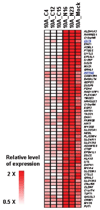
Concurrently down-regulated genes in MCF10A cells exposed to cancer–associated fibroblasts (CAFs). The 56 genes, harboring CpG islands, are shown in heat map. After co-cultured with CAFs, MCF10A cells (10A_C4, 10A_C12, and 10A_C15) were subjected to RNA extraction followed by gene expression analysis using the Affymetrix U133 plus 2.0 system. Expression profiling was also conducted in MCF10A cells (10A_N16 and 10A_N23) co-cultured with normal fibroblasts and in a control (10A_Mock) not exposed to fibroblasts. An additional list of 109 down-regulated genes, including those shown in the heatmap (n=56), is provided in Supplementary Table S2.
Figure 3
Figure 3
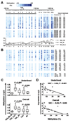
Methylation mapping and gene expression analyses of the CST6 CpG island in co-cultured MCF10A cells. A, twenty co-cultured MCF10A samples were subjected to the MassARRAY analysis as described in the text. Top: A genome map showing the locations of CpG sites and the transcription start site (TSS) of CST6. Middle: A methylation map derived from the MassARRAY analysis. Note that this assay will analyze multiple CpG dinucleotides together as a group if the sites are situated in close vicinity and within a digested fragment. Names of co-cultured samples and the average methylation levels of either the first 12 CpG units (underlined) or all 20 sites (overall) are shown at the right. Bottom: The landscape plots reveal greater levels of methylation in MCF10A cells co-cultured with cancer-associated fibroblasts (10A_CAF) than in cells co-cultured with normal fibroblasts (10A_NF) or a mock control (10A_Mock). B, upper and middle panels: Methylation levels of the CST6 CpG island in 20 monotypic fibroblasts (without MCF10A cells) were quantified. Lower panel: MassARRAY was used to assess the methylation levels of the CST6 CpG in MCF10A cells after the exposure to conditioned media obtained from cancer-associated (C8 CM) or from normal (N26 CM) fibroblast culture. C, box-plots summarize the methylation level of the overall (low) or the first 12 CpG units (upper) in MCF10A_Mock control, co-cultured MCF10A cells and monotypic breast fibroblasts. D, inverse correlation between methylation and expression levels of CST6. The MCF10A_Mock sample was plotted as the cross in the figures.
Figure 4
Figure 4
Epigenetic silencing of CST6 was induced by activated AKT1 signaling. A, positive correlation between the expression of CST6 and INPP4B in MCF10A cells co-cultured with various breast fibroblasts was shown. Logistic regression analysis was performed and spearman rank coefficient (SRC) was calculated. B, the co-cultured MCF10A cells were fixed and dually immunostained with anti-phospho-AKT1 (Texas-Red) and anti-epithelial specific antigen (FITC, green) followed by a nuclear staining with DAPI (blue). Representative images from confocal cross sections are shown. C, influence of cancer-associated fibroblasts on MCF10A cells was assessed by elevated phospho-AKT1 kinase in the latter cells. Fibroblasts and MCF10A cells were mixed in various ratios (shown in the x-axis) and grown on the Matrigel-coated chamber slides. Two weeks later, dual immunofluorescence (IF) staining was carried out, and the resultant mages were captured and analyzed as described in the text. The basal level of phospho-AKT1 kinase (red) signals detected in the mock control experiments was arbitrarily defined as 1. An average value of 15 images with +/− SD from 3 independent assessments is shown for each co-culture set. D, MCF10A cells were transfected with either an empty vehicle (vector ctrl) or with pCDNA3 encoding a constitutively active myristylated form of AKT1. Upper, AKT1 kinase activities in transfectants and in a positive control cell line (MDA-MB468) were measured in the absence (black bars) or presence (white bars) of an AKT1 kinase inhibitor. Data represented an average of 3 independent AKT1 kinase assessments. Lower, Expression levels of AKT1, CST6 and INPP4B in transfectants were measured by quantitative RT-PCR.
Figure 5
Figure 5
Repressive chromatin marks enriched at the CST6 locus. A, quantitative chromatin Immunoprecipitation (ChIP)-PCR analysis in AKT1 transfected and control MCF10A cells was shown. Enrichment levels of 3 histone marks, trimethyl-H3K27, dimetyl-H3K9, and acetyl-H3K9 and DNA methyltransferase 1 (DNMT1) were analyzed in the CST6 promoter and its surrounding regions (~15-kb). Data represented the average of 2 independent experiments. B, methylation analysis of the CST6 CpG by the MassARRAY analysis in AKT1-transfected MCF10A and vector control cells. Representative data were derived from at least 2 independent transfectants.
Figure 6
Figure 6
Methylation analysis of CST6 and immunostaining of phospho-AKT1 in primary breast tumors. A, methylation profiles of 194 primary breast tumors (upper) and 28 normal breast tissues (lower) were shown. The MassARRAY analysis was used to determine the methylation level of each sample. The methylation difference between cancer and normal tissues was determined to be significant in the overall (p <10−6) or the underlined (p <10−6) region (t-test). B, representative examples of differential expression of phospho-AKT on breast cancer tissue sections (Case 1: weak or undetectable with score 0; Case 2: strong with score 4, see Experimental Procedures for explanation). Arrows indicate tumor stromal cells that are in close contact with cancer epithelia. C, dot-plots indicate that the level of CST6 promoter methylation is positively correlated with the phospho-AKT1 staining intensity in 72 primary tumors available for analyses. The horizontal bars indicate mean values. Significance of differences in methylation was determined by student t-test.

References

    1. Callinan PA, Feinberg AP. The emerging science of epigenomics. Hum Mol Genet. 2006;1:95–101. - PubMed
    1. Fuks F. DNA methylation and histone modifications: teaming up to silence genes. Curr Opin Genet Dev. 2005;15:490–495. - VSports注册入口 - PubMed
    1. Antequera F, Bird A. CpG islands as genomic footprints of promoters that are associated with replication origins. Curr Biol. 1999;9:661–667. - "V体育官网" PubMed
    1. Jones PA, Baylin SB. The epigenomics of cancer. Cell. 2007;128:683–692. - V体育官网入口 - PMC - PubMed
    1. Plimack ER, Stewart DJ, Issa JP. Combining epigenetic and cytotoxic therapy in the treatment of solid tumors. J Clin Oncol. 2007;25:4519–4521. - PubMed

Publication types

MeSH terms