Skip to main page content
U.S. flag

An official website of the United States government

Dot gov

The . gov means it’s official. Federal government websites often end in . gov or . mil. Before sharing sensitive information, make sure you’re on a federal government site VSports app下载. .

Https

The site is secure V体育官网. The https:// ensures that you are connecting to the official website and that any information you provide is encrypted and transmitted securely. .

. 2009 Jan;119(1):20-32.
doi: 10.1172/JCI36226. Epub 2008 Dec 1.

Apoptotic human cells inhibit migration of granulocytes via release of lactoferrin

Affiliations

Apoptotic human cells inhibit migration of granulocytes via release of lactoferrin (V体育安卓版)

Irini Bournazou et al. J Clin Invest. 2009 Jan.

"V体育安卓版" Abstract

Apoptosis is a noninflammatory, programmed form of cell death. One mechanism underlying the non-phlogistic nature of the apoptosis program is the swift phagocytosis of the dying cells. How apoptotic cells attract mononuclear phagocytes and not granulocytes, the professional phagocytes that accumulate at sites of inflammation, has not been determined. Here, we show that apoptotic human cell lines of diverse lineages synthesize and secrete lactoferrin, a pleiotropic glycoprotein with known antiinflammatory properties. We further demonstrated that lactoferrin selectively inhibited migration of granulocytes but not mononuclear phagocytes, both in vitro and in vivo. Finally, we were able to attribute this antiinflammatory function of lactoferrin to its effects on granulocyte signaling pathways that regulate cell adhesion and motility. Together, our results identify lactoferrin as an antiinflammatory component of the apoptosis milieu and define what we believe to be a novel antiinflammatory property of lactoferrin: the ability to function as a negative regulator of granulocyte migration VSports手机版. .


Figures

Figure 1
Figure 1. Apoptotic cells release factor(s) that inhibit neutrophil migration.
(A) Immunohistochemical analysis of neutrophils in BL (left) and spleen (positive control; right) sections. Inset images represent isotype control. (B) Representative images of stained Transwell filters. (C) Neutrophil chemotaxis toward increasing concentrations of BL cells was assessed in the presence of fMLP (100 nM). n = 3; *P < 0.05 vs. time 0. (D) BL cell–conditioned media obtained at the indicated time points were used to analyze fMLP-induced neutrophil chemotaxis. n = 3; *P < 0.05 vs. fMLP. (E) Neutrophil chemotaxis toward fMLP was analyzed in the presence of control or Bcl-2–transfected BL2 cells obtained following a 0- and 5-hour incubation at 37°C. n = 3; *P < 0.05 vs. BL2 0 h; NS vs. BL2/Bcl-2 0 h. Apoptosis levels were assessed by flow cytometry following staining with annexin V/propidium iodide. Error bars indicate SEM. Original magnification; ×400 (A; A, insets; B). hpf, high-power field.
Figure 2
Figure 2. Biochemical characterization of the inhibitory factor.
Conditioned media from BL2 cells cultured for 24 hours were size fractionated using filters with 50 kDa (A) molecular weight cutoff sizes. Unfiltered medium was included as control. *P < 0.001 compared with the corresponding positive control. Error bars indicate SEM. Ion exchange analysis included the use of Q Sepharose beads (positively charged) in order to distinguish positively and negatively charged molecules in the <100 kDa fraction (B) of the BL medium. Unbound molecules (Q1 fraction) were collected, whereas bound molecules were eluted from the beads (Q2 fraction). Neutrophil migration toward these fractions in the presence of fMLP (100 nM) was assessed. Q1 and Q2 fractions (unbound and eluant fraction) of serum-free medium (no BL) were included as control. P < 0.05 compared with the corresponding control. Error bars indicate SEM. (C) Chemotaxis assay of neutrophils toward BL-conditioned medium that was heat inactivated (90°C for 10 minutes). (D) MALDI-TOF mass spectrum for the tryptic digest of the peptide band identified as lactoferrin.
Figure 3
Figure 3. Lactoferrin specifically inhibits neutrophil chemotaxis.
Neutrophil chemotaxis in the presence of human anti-lactoferrin (anti-LTF) polyclonal antibody (gray) or isotype control (black) using conditioned media from BL (A) and MCF7 (B) cells (A: n = 3, *P < 0.05 vs. isotype control, NS vs. fMLP anti-lactoferrin control; B; n = 3, P < 0.001 vs. fMLP/isotype; NS vs. fMLP/anti-LTF. (C) RT-PCR analysis to assess lactoferrin expression in BL cells stably expressing LTF shRNA (LTF) cells and mock-transfected (mm) cells induced to become apoptotic (1 μM staurosporine; 37°C). (D) Chemotaxis assay to determine neutrophil migration toward supernatants obtained from control, LTF shRNA, and mock-transfected BL cells (n = 5; §P < 0.05 compared with mm shRNA control; **P < 0.05 compared with fMLP; NS compared with fMLP control). (E) Dose-response analysis of purified human lactoferrin. n = 3; P < 0.05 vs. 0 g/ml purified LTF + fMLP. Error bars indicate SEM.
Figure 4
Figure 4. Neutrophil chemotaxis toward lactoferrin is irrespective of the chemoattractant used and its iron saturation status.
(A) Neutrophil chemotaxis toward different chemoattractants. n = 3; *P < 0.05. (B) Neutrophil chemotaxis toward chemoattractants (control) or chemoattractants that were incubated with lactoferrin (10 μg/ml) followed by the addition of isotype or anti-lactoferrin monoclonal antibody (10 μg/ml). Antibodies were removed using magnetic IgG beads. n = 3; *P < 0.05, NS compared with chemoattractant control. (C) Immunoblot analysis of lysates of neutrophils incubated with or without biotinylated lactoferrin (10 μg/ml) at 37°C for 1 hour. (D) Neutrophil chemotaxis toward lactoferrin (10 μg/ml) purified from human neutrophils or human milk. **P < 0.001 vs. fMLP. C5a-induced monocyte (E) or macrophage (F) chemotaxis. (G) Neutrophil migration in the presence of lactoferrin (10 μg/ml) in the top or bottom compartment of the Transwell insert (n = 3; NS vs. corresponding +LTF controls). (H) Chemotaxis assay to determine neutrophil migration toward purified recombinant iron-depleted (Apo-), partially iron-saturated, and fully iron-saturated (Holo-) recombinant lactoferrin (10 μg/ml). Milk-purified lactoferrin and partially iron-saturated transferrin (TF; 10 μg/ml) were added as control. n = 4; P < 0.001 compared with fMLP control. Error bars indicate SEM.
Figure 5
Figure 5. Lactoferrin inhibits neutrophil migration in vivo.
Total cell (A) or neutrophil number (Gr-1+; B) obtained from peritoneal lavage. *P < 0.05 vs. transferrin; P < 0.05 vs. thioglycollate (TG) control, **P < 0.01 vs. transferrin control. Error bars indicate SEM. (C) Characteristic cytospin images. Original magnification, ×400, top; ×200, bottom.
Figure 6
Figure 6. Effect of lactoferrin on neutrophil polarization morphology and spreading.
(A) Time-lapse video microscopy frames of control or lactoferrin-pretreated neutrophils (10 μg/ml; 40 minutes at 37°C) stimulated with 1 μM fMLP over a 1-hour incubation time course. Original magnification, ×400. (B) Quantification of neutrophils (nonpolarized) counted from 5 different fields; *P < 0.05, **P < 0.01 vs. corresponding +LTF control. Error bars indicate SEM. (C) Representative plot (of 3 independent experiments) showing measurement of [Ca2+]i levels in neutrophils incubated in the presence or absence of lactoferrin (10 μg/ml; 30 minutes at 37°C) followed by stimulation with 10 nM fMLP.
Figure 8
Figure 8. Induction of apoptosis upregulates lactoferrin expression and release.
(A) RT-PCR analysis in cell lines stimulated to undergo apoptosis (A) and unstimulated controls (V). MCF7 cells transfected with caspase-3 (25.4% apoptosis; 100 μM etoposide, 20 hours), Jurkat (18.4% apoptosis; 1 μM staurosporine, 3 hours), BL2 (12.46% apoptosis), and BL2/Bcl-2 (7.42% apoptosis; 1 μM staurosporine, 1 hour). The lanes were run on the same gel but, where indicated by the vertical lines, were noncontiguous. (B) Lactoferrin expression in A549 cells at defined time points (hours) following stimulation with 100 μM etoposide or 1 μM staurosporine. (C) Addition of pan-caspase inhibitor zVAD-fmk (100 μg/ml) for 12 hours in order to prevent etoposide-induced apoptosis in A549 cells. (D) Immunoblot analysis of cell supernatants from: BL2 and primary lymphocytes in the presence (+) or absence (–) of staurosporine (1 μM) in serum-free conditions for 1 hour. A549 cells were stimulated with (+) or without (–) 100 μM etoposide for 5 hours. All samples were run on the same gel. Noncontiguous samples of A549 cells and lymphocytes (Lymph) are indicated by the vertical lines. (E) A549 cells were induced to become apoptotic (100 μM etoposide; 20 hours) in the presence or absence of brefeldin A (1 μg/ml), a protein release inhibitor. (F) Immunoblot analysis of cell supernatants from control BL2 cells (1 × 10⁶/ml) induced to undergo apoptosis (1 μM staurosporine, 1 hour) or primary necrosis (56°C, 1 hour) in serum-free conditions. St, staurosporine; con; control; Etop, etoposide.
Figure 7
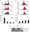
Figure 7. Effect of lactoferrin on neutrophil activation status.
The expression of CD62L (A and B) and CD11b (C and D) was assessed in fMLP- (100 nM), TNF-α– (1 ng ml), or PMA-stimulated (100 nM) neutrophils (30 minutes at 37°C) that were preincubated (40 minutes at 37°C) in the presence or absence of lactoferrin (10 μg/ml). Representative flow cytometry overlays of CD62L (A) and CD11b (C) expression in control (gray) and stimulated neutrophils (lactoferrin-treated: red; untreated: blue). n = 3; *P < 0.05, **P < 0.01. Error bars indicate SEM. (E) Western blot analysis to determine levels of ERK1/2 phosphorylation. Neutrophils were incubated with lactoferrin (10 μg/ml; 40 minutes at 37°C), followed by stimulation with fMLP (100 nM) for the indicated times. Membrane was stripped and reprobed for total ERK2. Results are representative of 3 independent experiments.

References

    1. Lauber K., et al. Apoptotic cells induce migration of phagocytes via caspase-3-mediated release of a lipid attraction signal. Cell. 2003;113:717–730. doi: 10.1016/S0092-8674(03)00422-7. - DOI - PubMed
    1. Truman L.A., et al. CX3CL1/fractalkine is released from apoptotic lymphocytes to stimulate macrophage chemotaxis. . Blood. 2008 doi: 10.1182/blood-2008-06-162404. - DOI - PubMed
    1. Henson P.M., Hume D.A. Apoptotic cell removal in development and tissue homeostasis. Trends Immunol. 2006;27:244–250. doi: 10.1016/j.it.2006.03.005. - DOI - PubMed
    1. Ravichandran K.S., Lorenz U. Engulfment of apoptotic cells: signals for a good meal. Nat. Rev. Immunol. 2007;7:964–974. doi: 10.1038/nri2214. - DOI - PubMed
    1. Savill J., Dransfield I., Gregory C., Haslett C. A blast from the past: clearance of apoptotic cells regulates immune responses. Nat. Rev. Immunol. 2002;2:965–975. doi: 10.1038/nri957. - DOI - PubMed

Publication types

MeSH terms