<b dir="3SEyUt1"></b> Skip to main page content
U.S. flag

An official website of the United States government

Dot gov

The . gov means it’s official. Federal government websites often end in VSports app下载. gov or . mil. Before sharing sensitive information, make sure you’re on a federal government site. .

Https

The site is secure. The https:// ensures that you are connecting to the official website and that any information you provide is encrypted and transmitted securely V体育官网. .

. 2008 Nov 15;68(22):9459-68.
doi: 10.1158/0008-5472.CAN-08-2634.

Regulated expression of a tumor-associated antigen reveals multiple levels of T-cell tolerance in a mouse model of lung cancer

Affiliations

Regulated expression of a tumor-associated antigen reveals multiple levels of T-cell tolerance in a mouse model of lung cancer

VSports注册入口 - Ann F Cheung et al. Cancer Res. .

Abstract

Maximizing the potential of cancer immunotherapy requires model systems that closely recapitulate human disease to study T-cell responses to tumor antigens and to test immunotherapeutic strategies. We have created a new system that is compatible with Cre-LoxP-regulatable mouse cancer models in which the SIY antigen is specifically overexpressed in tumors, mimicking clinically relevant TAAs. To show the utility of this system, we have characterized SIY-reactive T cells in the context of lung adenocarcinoma, revealing multiple levels of antigen-specific T-cell tolerance that serve to limit an effective antitumor response. Thymic deletion reduced the number of SIY-reactive T cells present in the animals. When potentially self-reactive T cells in the periphery were activated, they were efficiently eliminated. Inhibition of apoptosis resulted in more persistent self-reactive T cells, but these cells became anergic to antigen stimulation VSports手机版. Finally, in the presence of tumors overexpressing SIY, SIY-specific T cells required a higher level of costimulation to achieve functional activation. This system represents a valuable tool in which to explore sources contributing to T-cell tolerance of cancer and to test therapies aimed at overcoming this tolerance. .


Figures

Figure 1
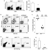
Figure 1. R26LSL-LSIY exhibits Cre-dependent over-expression
A. Luciferase activity in R26+/+ and R26LSL-LSIY/+ MEFs uninfected (n = 2 and 12, open bars) or infected with Ad-Cre (n = 3 and 12, filled bars). Each bar shows mean + s.e.m with Student’s t-tests: p < 0.0001 for both R26LSL-LSIY/+ -Cre versus +Cre and -Cre R26+/+ versus R26LSL-LSIY/+ B. Left panel: Number of CD8+1B2+ cells in tail tip bleeds of R26+/+;2C and R26LSL-LSIY/+;2C mice. Each bar shows mean + s.e.m. with p < 0.0001 by Student’s t-test. Right panel: 1B2, CD4, and CD8 staining of thymocytes from representative R26+/+;2C (top) and R26LSL-LSIY/+;2C (bottom) mice. C. Luciferase activity in tissue lysates from whole thymus, lung, liver, and spleen from R26+/+ (n = 4, open bars), R26LSL-LSIY/+ (n = 5, shaded gray bars), and R261Lox-LSIY/+ (n = 2, filled black bars) mice. Each bar shows mean + s.e.m. with *p < 0.05, **p < 0.005, ***p ≪ 0.001 by Student’s t-test. D. Representative CFSE dilution in CD8+1B2+cells recovered from mesenteric lymph nodes and spleens of R26+/+ (filled histograms) and R26LSL-LSIY/+ (open histograms) recipients of naïve 2C cells one, two, and three days after i.v. transfer.
Figure 2
Figure 2. Central tolerance to SIY is incomplete and functional SIY-reactive T cells can be transiently activated in the periphery
A. Left panel: Representative plots of CD8 and BD Dimer X SIY/H-2Kb:Ig stained whole lung cell suspensions from R26+/+ and R26LSL-LSIY/+ mice seven days after intranasal WSN-SIY Influenza A infection. Right panel: SIY/H-2Kb:Ig+ fraction of total CD8+ cells from WSN-SIY-infected lungs of R26+/+ (n = 4, open bar) and R26LSL-LSIY/+ (n = 4, filled bar) mice. Each bar shows mean + s.e.m. with p < 0.005 by Student’s t-test. B. Representative plots of single cell suspensions from lungs (top panels) and mediastinal lymph nodes (bottom panels) of R26+/+ and R26LSL-LSIY/+ mice 7 days post-WSN-SIY infection and uninfected controls. Cells were cultured and stimulated in vitro with 1ug/mL of SIY peptide and an IFN-γ capture assay was performed. CD8+-gated cells are stained for CD107a and secreted IFN-γ. C. SIY-specific cytotoxicity in mediastinal lymph nodes of R26+/+ and R26LSL-LSIY/+ mice 7 days after pulmonary WSN-SIY vaccination. Mean +/− s.e.m. specific cytolysis are shown for each genotypes. D. Left panel: BD DimerX SIY/H-2Kb:Ig double-stained splenocytes from mice boosted with DC2.4.LSIY vaccine 5 days prior to analysis. R26+/+ (left) and R26LSL-LSIY/+ (middle) mice were infected intranasally with WSN-SIY >30 days before boost or never infected (right). Right panel: Quantification of percentage of double-positive SIY/H-2Kb:Ig cells among splenocytes in R26+/+ (n = 4, open bar) and R26LSL-LSIY/+ (n = 3, filled bar) mice. Each bar shows mean + s.e.m. with p < 0.05 by Student’s t-test.
Figure 3
Figure 3. Antigen over-expressing tumors progress normally
A. Representative H&E-staining lung tumor histology from K-rasLSL-G12D/+;R26+/+ (left) and K-rasLSL-G12D/+;R26LSL-LSIY/+ (right) mice 12 weeks after intranasal Ad-Cre infection. Scale bars indicate 100μm. B. Luciferase activity of representative individual lung tumors dissected from K-rasLSL-G12D/+;R26+/+ (blue bars) and K-rasLSL-G12D/+;R26LSL-LSIY/+ (red bars) animals 16 weeks after intranasal Ad-Cre infection. C. In vivo bioluminescence of a K-rasLSL-G12D/+;R26LSL-LSIY/+ mouse > 6 months after intranasal Ad-Cre infection (inset: gross image of lungs after imaging). D. Quantification of lung tumor burden in K-rasLSL-G12D/+;R26+/+ (n = 3, blue bars) and K-rasLSL-G12D/+;R26LSL-LSIY/+ (n = 3, red bars) animals 12 weeks after intranasal Ad-Cre infection. Each bar shows mean + s.e.m. with p = 0.89 and 0.83 for tumor to lung ratio and average tumor area by Student’s t-test.
Figure 4
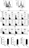
Figure 4. Naïve cells recognize, but do not response effectively to lung tumors
A. Representative plot of CD44 staining in CD8+1B2+ cells 5 days after naïve 2C cell transfer into K-rasLSL-G12D/+;R26+/+ (filled histogram) and K-rasLSL-G12D/+;R26LSL-LSIY/+ (open histogram) animals. B. Representative plot of CFSE dilution in CD8+1B2+ cells 3 days after naïve 2C cell transfer into K-rasLSL-G12D/+;R26+/+ (filled histogram) and K-rasLSL-G12D/+;R26LSL-LSIY/+ (open histogram) animals. C. Top panel: Representative plots of 1B2 and BD Dimer X SIY/H-2Kb:Ig stained cells recovered from lymph nodes of tumor-free and tumor-bearing R26+/+ and R26LSL-LSIY/+ mice. The ratio of 1B2+SIY/H-2Kb:Ig+ cells in mediastinal to mesenteric lymph nodes is indicated. Double staining with 1B2 and Dimer X to detect 2C cells enhances the signal to noise ratio. Bottom panel: Representative plots of whole lung cell suspensions from the same tumor-free and tumor-bearing R26+/+ and R26LSL-LSIY/+ mice stained with 1B2 and BD Dimer X SIY/H-2Kb:Ig. D. Tumor to lung area ratio in K-rasLSL-G12D/+;R26+/+ (n = 9, open bars) and K-rasLSL-G12D/+;R26LSL-LSIY/+ (n = 8, filled bars) animals 16 weeks after intranasal Ad-Cre infection, shown in left graft. Animals received naïve 2C cell i.v. transfer at either 4 weeks or 12 weeks after intranasal Ad-Cre. Each bar shows mean + s.e.m. with p = 0.060 by Student’s t-test for tumor burden. The average area of tumors (p = 0.046 by Student’s t-test) in the same animals are shown in the middle graph and the average number of tumors per animal (p = 0.096 by Student’s t-test) is shown in the right graph.
Figure 5
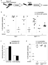
Figure 5. A higher threshold for costimulation is required in the presence of tumors over-expressing self-antigen
A. Schematic of experimental set-up for the in vivo cytotoxicity assay. Naïve 2C cells are intravenously injected into recipients with or without intraperitoneal DC2.4.LSIY or intranasal WSN-SIY vaccination. Six days later, differentially-labeled R261LoxLSIY/+ target and R26+/+ control cells are intravenously injected and animals are analyzed 20–24 hours later. B. SIY-specific cytotoxicity in mediastinal lymph nodes draining the lung seven days after naïve 2C cell transfer into recipients with (closed circles) or without (open circles) DC2.4.LSIY vaccination. C. Fold change in SIY-specific cytotoxicity in mediastinal (open bars) and mesenteric lymph nodes (filled bars) of tumor-free and tumor-bearing R26LSL-LSIY/+ mice when vaccinated with DC2.4.LSIY over unvaccinated mice. D. SIY-specific cytotoxicity in mediastinal lymph nodes draining the lung seven days after naïve 2C cell transfer into recipients with (closed circles) or without (open circles) WSN-SIY vaccination
Figure 6
Figure 6. Inhibition of apoptosis results in T cell anergy in the presence of self-antigen
A. In vitro activated 2C cells infected with MIG-Bcl2 propagated in vitro in the presence (filled circles) or absence (open circles) of survival cytokine IL-2. CD8+1B2+ cells were analyzed by flow cytometry for the percentage of EGFP+ cells each day for 4 days after withdrawal of IL-2. B. In vitro activated 2C cells infected with MIG-Bcl2 intravenously transferred into either R26+/+ (open circles) or R26LSL-LSIY/+ (filled circles) recipient mice. Spleens were harvested and analyzed by flow cytometry at 2, 4, 8, and 16 days after transfer. EGFP+ cells as a percentage of total CD8+1B2+ (top panel) and CD8+1B2+ cells as a percentage of total splenocytes (bottom panel) are shown. C. Representative plots of CD8+-gated splenocytes from R26+/+ and R26LSL-LSIY/+ mice 8 days after receiving MIG-Bcl2 infected activated 2C cells. Cells were stimulated in vitro with 1ug/mL of SIY peptide (right panels) or unstimulated (left panels) and an IFN-γ capture assay was performed. Population gates represent percentage of EGFP IFN-γsecreting (gray gate value) and EGFP+ IFN-γ-secreting (black gate value) CD8+ splenocytes.

References

    1. Dunn GP, Old LJ, Schreiber RD. The three Es of cancer immunoediting. Annu Rev Immunol. 2004;22:329–60. - V体育官网 - PubMed
    1. Rabinovich GA, Gabrilovich D, Sotomayor EM. Immunosuppressive strategies that are mediated by tumor cells. Annu Rev Immunol. 2007;25:267–96. - PMC - PubMed
    1. Swann JB, Smyth MJ. Immune surveillance of tumors. J Clin Invest. 2007;117(5):1137–46. - PMC - PubMed
    1. Anderson MJ, Shafer-Weaver K, Greenberg NM, Hurwitz AA. Tolerization of tumor-specific T cells despite efficient initial priming in a primary murine model of prostate cancer. J Immunol. 2007;178(3):1268–76. - PubMed
    1. Drake CG, Doody AD, Mihalyo MA, et al. Androgen ablation mitigates tolerance to a prostate/prostate cancer-restricted antigen. Cancer Cell. 2005;7(3):239–49. - "V体育2025版" PMC - PubMed

Publication types

"VSports" LinkOut - more resources