Skip to main page content
U.S. flag

An official website of the United States government

Dot gov

The . gov means it’s official. Federal government websites often end in . gov or . mil. Before sharing sensitive information, make sure you’re on a federal government site VSports app下载. .

Https

The site is secure. The https:// ensures that you are connecting to the official website and that any information you provide is encrypted and transmitted securely V体育官网. .

. 2008 Nov 28;283(48):33437-46.
doi: 10.1074/jbc.M802016200. Epub 2008 Oct 1.

HMGA2 and Smads co-regulate SNAIL1 expression during induction of epithelial-to-mesenchymal transition

Affiliations

HMGA2 and Smads co-regulate SNAIL1 expression during induction of epithelial-to-mesenchymal transition (VSports)

Sylvie Thuault et al. J Biol Chem. .

Abstract

Epithelial-mesenchymal transition (EMT) is important during embryonic cell layer movement and tumor cell invasiveness. EMT converts adherent epithelial cells to motile mesenchymal cells, favoring metastasis in the context of cancer progression. Transforming growth factor-beta (TGF-beta) triggers EMT via intracellular Smad transducers and other signaling proteins. We previously reported that the high mobility group A2 (HMGA2) gene is required for TGF-beta to elicit EMT in mammary epithelial cells. In the present study we investigated the molecular mechanisms by which HMGA2 induces EMT VSports手机版. We found that HMGA2 regulates expression of many important repressors of E-cadherin. Among these, we analyzed in detail the zinc-finger transcription factor SNAIL1, which plays key roles in tumor progression and EMT. We demonstrate that HMGA2 directly binds to the SNAIL1 promoter and acts as a transcriptional regulator of SNAIL1 expression. Furthermore, we observed that HMGA2 cooperates with the TGF-beta/Smad pathway in regulating SNAIL1 gene expression. The mechanism behind this cooperation involves physical interaction between these factors, leading to an increased binding of Smads to the SNAIL1 promoter. SNAIL1 seems to play the role of a master effector downstream of HMGA2 for induction of EMT, as SNAIL1 knock-down partially reverts HMGA2-induced loss of epithelial differentiation. The data propose that HMGA2 acts in a gene-specific manner to orchestrate the transcriptional network necessary for the EMT program. .


Figures (V体育安卓版)
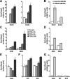
FIGURE 1.
FIGURE 1.
HMGA2 regulates different E-cadherin transcriptional repressors. Expression levels of mRNA for Snail1 and Snail2 (A), ZEB1 and ZEB2 (C), Twist (E), and E47 and E2-2 (F) was assessed by quantitative RT-PCR in NMuMG cells treated or not with 5 ng/ml TGF-β1 for 2, 8, and 24 h. Expression levels of mRNAs for Snail1 and Snail2 (B), ZEB1 and ZEB2 (D), Twist, E47, and E2-2 (G) was assessed by quantitative RT-PCR in parental NMuMG or in a cell clone of NMuMG expressing constitutively human HMGA2 (HMGA2-NMuMG). Average values are plotted in the bar graphs with S.D. derived from triplicate determinations.
FIGURE 2.
FIGURE 2.
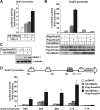
HMGA2 and Smad3/Smad4 cooperate to activate the Snail1 promoter. A, luciferase reporter assays of the Snail1 promoter construct in HepG2 cells transiently transfected (+) or not (-) with an HA-HMGA2 expression construct and treated (+) or not (-) with 1 ng/ml TGF-β1 for 24 h. B, luciferase reporter assays of the Snail1 promoter construct in HepG2 cells transiently transfected with FLAG-Smad3, FLAG-Smad4, and HA-HMGA2 expression constructs as indicated. Levels of expression of the transfected proteins were assessed by immunoblot using anti-FLAG and anti-HA antibodies. Stars indicate nonspecific bands. C, immunoblot analysis of endogenous SNAIL1 in HepG2 cells transiently transfected with FLAG-Smad3, FLAG-Smad4, and HA-HMGA2 expression constructs as indicated. α-Tubulin served as a loading control. D, luciferase reporter assays of the indicated deletion constructs of the Snail1 promoter in HepG2 cells transiently transfected with FLAG-Smad3, FLAG-Smad4, and HA-HMGA2 expression constructs as indicated. A schematic representation of potential binding elements for different transcription factors present in the Snail1 promoter and the breakpoints of the deletion constructs used are shown. In all panels normalized luciferase data are plotted in bar graphs as averages with S.D. derived from triplicate determinations.
FIGURE 3.
FIGURE 3.
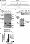
HMGA2 binds and increases binding of Smad3 to the Snail1 promoter. A, nucleotide sequences of wild type (wt) and mutant (m1–4) Snail1 promoter probes used for the DNAP experiments. Nucleotide numbers were assigned relative to the ATG codon. A line indicates that the sequence has not been altered. B, binding of HMGA2 (HA-HMGA2) and C-terminal-phosphorylated Smad3 (P-Smad3) to wild type Snail1 promoter probes described in panel A was assessed by DNAP experiments using extracts of transiently transfected HepG2 cells with FLAG-Smad3, FLAG-Smad4, and HA-HMGA2 expression constructs treated (+) or not (-) with 5 ng/ml TGF-β1 for 2 h. The probes used are indicated on the left. TCL, total cell lysates. WB, Western blot. C, binding of HMGA2 to wild type and mutant (m1, m2) Snail1 promoter probes spanning from -131 to -92 described in panel A was assessed by DNAP experiments using extracts of transiently transfected HepG2 cells with the HA-HMGA2 expression construct. D, luciferase reporter assays of wild type or mutant (m1) -625 Snail1 promoter constructs in HepG2 cells transiently transfected with HA-HMGA2 and/or FLAG-Smad3 and FLAG-Smad4 expression constructs as indicated. E, binding of endogenous Smad4 to the Snail1 promoter was assessed by chromatin immunoprecipitation (IP) of NMuMG cells treated (+) or not (-) with 5 ng/ml TGF-β1 for 2 h. ctrl, control. F, binding of phosphorylated Smad3 (P-Smad3) to wild type and mutant (m3, m4) probes spanning -230 to -178 of the Snail1 promoter described in panel A was assessed by DNAP experiments using extracts of HepG2 cells described in panel B.
FIGURE 4.
FIGURE 4.
HMGA2 interacts with Smads. A, pulldown of 293T cell extracts transfected with FLAG-Smad2, -Smad3, or -Smad4 with purified GST or GST-HMGA2. The input transfected proteins are shown in an immunoblot of the total cell lysate (TCL). WB, Western blot. B, binding of endogenous Smad3 from HepG2 cell extracts treated (+) or not (-) with 2.5 ng/ml TGF-β1 for 2 h to GST or GST-HMGA2 was assessed. C, pulldown of in vitro translated Smad3 and Smad4 with purified GST or GST-HMGA2. Autoradiograms of the bound proteins and 2% of the input of in vitro translated proteins are shown. D, pulldown of 293T cell extracts transfected with domains of 6×Myc-Smad3 as indicated with purified GST or GST-HMGA2. FL, full-length. E, pulldown assay of HepG2 cell extracts with purified GST or deletion mutants of GST-HMGA2. HMGA2 deletion mutants are schematically depicted, and their ability to interact with Smad3 is summarized with +/-. Ponceau staining visualizes the input GST fusion proteins used in the pulldown.
FIGURE 5.
FIGURE 5.
The acidic C-terminal domain of HMGA2 is required for Snail1 promoter activation. A, luciferase reporter assays of the Snail1 promoter construct in COS1 cells transiently transfected with constructs for HA-HMGA2 wild type (wt) or HA-HMGA2 with its acidic C-terminal domain deleted (ΔC). Luciferase data are plotted as in Fig. 2, and immunoblot of the transfected HMGA2 proteins is shown below the graph. B, binding of wt or ΔC HMGA2 to the Snail1 promoter probe -131/-92 was assessed by DNAP experiments in COS1 cells. DNA binding specificity is shown using the Smad-specific 4× CAGA DNA probe. WB, Western blot; TCL, total cell lysates.
FIGURE 6.
FIGURE 6.
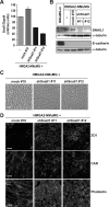
Snai11 depletion partially reverts the mesenchymal phenotype associated with ectopic HMGA2 expression. A, analysis of Snail1 expression by quantitative RT-PCR in HMGA2-NMuMG cells non-transfected (-) and stably transfected with the empty vector (mock #10) or with a construct expressing a short hairpin RNA against Snail1 (shSnail1#11 and shSnail1#12, #11 and #12 being two independent clones). B, analysis of Snail1 and E-cadherin protein levels in cell clones described in panel A. α-Tubulin served as a loading control. NMuMG-m corresponds to a sub-clone of NMuMG stably transfected with the empty pMEP4 vector exhibiting a highly polarized epithelial phenotype. C, phase-contrast microscopy of cells stably transfected with the specific shRNA expressing vectors described in panel A. D, visualization of the epithelial tight junction markers ZO-1 and CAR and of the actin cytoskeleton by immunostaining of the cells stably transfected with the specific shRNA expressing vectors described in panel A. Bars represent 10 μm.
FIGURE 7.
FIGURE 7.
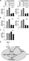
Snail1 depletion decreases expression of specific repressors of E-cadherin induced by HMGA2. Expression levels of Snail1 and Snail2 (A), ZEB1 and ZEB2 (B), and Twist (C) were assessed by quantitative RT-PCR in cells described in Fig. 6. The data are plotted as described in Fig. 1. D, model of the molecular mechanism by which TGF-β via Smads induces HMGA2 that then, in collaboration with the Smads, regulates SNAIL1 expression, which in turn regulates additional repressors of E-cadherin to induce EMT. The connection between HMGA2 and TWIST remains unclear (question mark).

References

    1. Thiery, J.-P., and Sleeman, J. P. (2006) Nat. Rev. Mol. Cell Biol. 7 131-142 - "V体育ios版" PubMed
    1. Peinado, H., Portillo, F., and Cano, A. (2004) Int. J. Dev. Biol. 48 365-375 - PubMed
    1. Peinado, H., Olmeda, D., and Cano, A. (2007) Nat. Rev. Cancer 7 415-428 - VSports最新版本 - PubMed
    1. Yang, J., Mani, S. A., Donaher, J. L., Ramaswamy, S., Itzykson, R. A., Come, C., Savagner, P., Gitelman, I., Richardson, A., and Weinberg, R. A. (2004) Cell 117 927-939 - PubMed
    1. Kondo, M., Cubillo, E., Tobiume, K., Shirakihara, T., Fukuda, N., Suzuki, H., Shimizu, K., Takehara, K., Cano, A., Saitoh, M., and Miyazono, K. (2004) Cell Death Differ. 11 1092-1101 - VSports最新版本 - PubMed

Publication types

V体育2025版 - MeSH terms