"VSports app下载" Skip to main page content
U.S. flag

An official website of the United States government

Dot gov

The . gov means it’s official VSports app下载. Federal government websites often end in . gov or . mil. Before sharing sensitive information, make sure you’re on a federal government site. .

Https

The site is secure. The https:// ensures that you are connecting to the official website and that any information you provide is encrypted and transmitted securely. V体育官网.

. 2009 Jan;24(1):50-61.
doi: 10.1359/jbmr.080817.

Mechanical stimulation of mesenchymal stem cell proliferation and differentiation promotes osteogenesis while preventing dietary-induced obesity

Affiliations

Mechanical stimulation of mesenchymal stem cell proliferation and differentiation promotes osteogenesis while preventing dietary-induced obesity (VSports注册入口)

Yen Kim Luu et al. J Bone Miner Res. 2009 Jan.

Abstract

Mesenchymal stem cells (MSCs) are defined by their ability to self-renew and differentiate into the cells that form mesodermal tissues such as bone and fat. Low magnitude mechanical signals (LMMS) have been shown to be anabolic to bone and have been recently reported to suppress the development of fat in normal animals fed a regular diet. Using male C57BL/6J mice, the ability of LMMS (0. 2g, 90-Hz signal applied for 15 min/d, 5 d/wk) to simultaneously promote bone formation and prevent diet-induced obesity was correlated to mechanical influences on the molecular environment of the bone marrow, as indicated by the population dynamics and lineage commitment of MSCs VSports手机版. Six weeks of LMMS increased the overall marrow-based stem cell population by 37% and the number of MSCs by 46%. Concomitant with the increase in stem cell number, the differentiation potential of MSCs in the bone marrow was biased toward osteoblastic and against adipogenic differentiation, as reflected by upregulation of the transcription factor Runx2 by 72% and downregulation of PPARgamma by 27%. The phenotypic impact of LMMS on MSC lineage determination was evident at 14 wk, where visceral adipose tissue formation was suppressed by 28%, whereas trabecular bone volume fraction in the tibia was increased by 11%. Translating this to the clinic, a 1-yr trial in young women (15-20 yr; n = 48) with osteopenia showed that LMMS increased trabecular bone in the spine and kept visceral fat at baseline levels, whereas control subjects showed no change in BMD, yet an increase in visceral fat. Mechanical modulation of stem cell proliferation and differentiation indicates a unique therapeutic target to aid in tissue regeneration and repair and may represent the basis of a nonpharmacologic strategy to simultaneously prevent obesity and osteoporosis. .


Figures

FIG. 1
FIG. 1
Representative density dot plots from flow cytometry experiments indicate the ability of LMMS to increase the number of stem cells in general (Sca-1 single positive, top quadrants), and MSCs specifically (both Sca-1 and Pref-1 positive, top right quadrant). Red, high cell density; blue, low cell density. Compared with control animals (A), LLMSs increase the number of stem cells in the bone marrow of LMMS animals (B). The actual increase in total bone marrow–derived stem cell number (C) and MSC number (D) was calculated as percent positive cells/total cells for the cell fraction showing highest intensity staining.
FIG. 2
FIG. 2
LMMS influence on stem cells was focused on the distinct cell populations identified in flow cytometry (A), with stem cells being identified as low forward (FSC) and side (SSC) scatter. Osteoprogenitor cells were identified as Sca-1+ cells, residing in the region highlighted as high FSC and SSC, and were 29.9% (p = 0.23) more abundant in the bone marrow of LMMS-treated animals (B). The pre-adipocyte population, identified as Pref-1+, Sca-1, showed a trend (+18.5%; p = 0.25) toward an increase in LMMS relative to CON animals (C).
FIG. 3
FIG. 3
Relative to CON, LMMS biases the bone marrow environment toward osteogenesis and away from adipogenesis. Real-time RT-PCR analysis of bone marrow samples harvested from animals subject to 6-wk LMMS treatment or sham control indicated a significant upregulation of the osteogenic gene Runx2 (A) and downregulation of the adipogenic gene PPARγ (B). Data are shown as expression levels relative to values for sham handled CON animals (represented as 1.0).
FIG. 4
FIG. 4
Bone volume fraction, as measured in vivo by low-resolution μCT, indicated that LMMS increased bone volume fraction across the entire torso of the animal (A). After death, high-resolution CT of the proximal tibia indicated a significant increase in trabecular bone density (B). Body mass of the animal at death was used as a covariate in the statistical analysis. Compared with controls (C), representative μCT reconstructions of the proximal tibia indicate the enhanced morphological properties of LMMS animals.
FIG. 5
FIG. 5
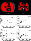
Representative in vivo μCT images used to discriminate visceral and subcutaneous adiposity in the abdominal region of a CON and LMMS animal. Visceral fat is shown in red; subcutaneous fat in gray (A). Linear regressions of calculated VAT volume against adipose and liver biochemistry values showed strong positive correlations in CON, and weak correlations in LMMS groups, as well as generally lower levels for all LMMS biochemical values (n = 6 for adipose, n = 10 for liver). Regressions for adipose TG (p = 0.002, B), adipose NEFA (p = 0.03, C), liver TG (p = 0.006, D), and liver NEFA (p = 0.003, E) were significant for CON animals, but only liver NEFA (p = 0.02) was significant for LMMS. Overall, LMMS mice exhibited lower, nonsignificant correlations in liver TG (p = 0.06), adipose TG (p = 0.19), and adipose NEFA (p = 0.37) to increases in visceral adiposity. ○, CON; ■, LMMS.
FIG. 6
FIG. 6
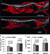
Suppression of the obese phenotype was achieved to a degree by stem cells preferentially diverting from an adipogenic lineage. Reconstructed in vivo μCT images of total body fat (red; A) indicate that, after 12 wk, animals that began LMMS at the time that the high-fat diet was introduced exhibited 22.2% less fat volume compared with controls. In contrast, animals allowed a high-fat diet for 4 wk before LMMS failed to show any reduction of fat volume (B). Shown as a relative percentage of fat to total animal volume, LMMS reduced the percent animal adiposity by 13.5% (p = 0.017), whereas the lack of a response in the already obese animals reinforces a conclusion that the mechanical signal works primarily at the stem cell development level, because existing fat is not metabolized by LMMS stimulation.
FIG. 7
FIG. 7
As measured by CT scans in the lumbar region of the spine, a group of young osteopenic women subject to LMMS for 12 mo (n = 24; gray bars ± SE) increased both BMD (p = 0.025 relative to baseline; mg/cm3) and muscle area (p < 0.001; cm2), changes that were paralleled by a nonsignificant increase in visceral fat formation (p = 0.45; cm2). Conversely, women in the CON group (n = 24; white bars ± SE), while failing to increase either BMD (p = 0.93) or muscle area (p = 0.43), realized a significant increase in visceral fat formation (p = 0.03). *Changes that are significantly different from baseline.

References

    1. Cheng SL, Shao JS, Charlton-Kachigian N, Loewy AP, Towler DA. MSX2 promotes osteogenesis and suppresses adipogenic differentiation of multipotent mesenchymal progenitors. J Biol Chem. 2003;278:45969–45977. - PubMed
    1. David V, Martin A, Lafage-Proust MH, Malaval L, Peyroche S, Jones DB, Vico L, Guignandon A. Mechanical loading down-regulates peroxisome proliferator-activated receptor gamma in bone marrow stromal cells and favors osteoblastogenesis at the expense of adipogenesis. Endocrinology. 2007;148:2553–2562. - PubMed (VSports注册入口)
    1. Gesta S, Tseng YH, Kahn CR. Developmental origin of fat: Tracking obesity to its source. Cell. 2007;131:242–256. - V体育平台登录 - PubMed
    1. Ghosh K, Ingber DE. Micromechanical control of cell and tissue development: Implications for tissue engineering. Adv Drug Deliv Rev. 2007;59:1309–1318. - V体育官网 - PubMed
    1. Rehfeldt F, Engler AJ, Eckhardt A, Ahmed F, Discher DE. Cell responses to the mechanochemical microenvironment-Implications for regenerative medicine and drug delivery. Adv Drug Deliv Rev. 2007;59:1329–1339. - PMC - PubMed

"VSports最新版本" Publication types