Skip to main page content
U.S. flag

An official website of the United States government

Dot gov

The . gov means it’s official. Federal government websites often end in . gov or . mil. Before sharing sensitive information, make sure you’re on a federal government site. VSports app下载.

Https

The site is secure V体育官网. The https:// ensures that you are connecting to the official website and that any information you provide is encrypted and transmitted securely. .

. 2008 Oct;28(19):5951-64.
doi: 10.1128/MCB.00305-08. Epub 2008 Aug 4.

A gene signature-based approach identifies mTOR as a regulator of p73

Affiliations

A gene signature-based approach identifies mTOR as a regulator of p73

Jennifer M Rosenbluth et al. Mol Cell Biol. 2008 Oct.

Abstract

Although genomic technologies have advanced the characterization of gene regulatory networks downstream of transcription factors, the identification of pathways upstream of these transcription factors has been more challenging. In this study we present a gene signature-based approach for connecting signaling pathways to transcription factors, as exemplified by p73. We generated a p73 gene signature by integrating whole-genome chromatin immunoprecipitation and expression profiling. The p73 signature was linked to corresponding signatures produced by drug candidates, using the in silico Connectivity Map resource, to identify drugs that would induce p73 activity. Of the pharmaceutical agents identified, there was enrichment for direct or indirect inhibitors of mammalian Target of Rapamycin (mTOR) signaling. Treatment of both primary cells and cancer cell lines with rapamycin, metformin, and pyrvinium resulted in an increase in p73 levels, as did RNA interference-mediated knockdown of mTOR. Further, a subset of genes associated with insulin response or autophagy exhibited mTOR-mediated, p73-dependent expression. Thus, downstream gene signatures can be used to identify upstream regulators of transcription factor activity, and in doing so, we identified a new link between mTOR, p73, and p73-regulated genes associated with autophagy and metabolic pathways VSports手机版. .


Figures
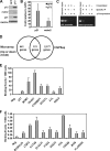
FIG. 1.
FIG. 1.
Generation of a multitiered p73 signature. H1299 cells were transduced with TAp73β- or GFP-expressing adenoviruses. (A) Protein lysates were harvested after transduction, and p73, GAPDH, and downstream targets mdm2 and p21 were analyzed by Western blotting. (B and C) p73 regulates known target genes when expressed in H1299 cells. (B) Total RNA was purified and reverse transcribed, and quantitative real-time PCR was performed with primers for p21 and mdm2. The samples were normalized to GAPDH, and the results are presented as increases over values for GFP control. Error bars represent standard deviations from three experiments. (C) For ChIP analysis, p73 was immunoprecipitated (IP) from formaldehyde-cross-linked H1299 cells transduced with adenoviral p73 or a GFP control. Associated DNA fragments were PCR amplified using primers flanking the p53 family response elements in p21 and mdm2. Nonspecific binding was assayed by immunoprecipitation (IP) of p73 from non-cross-linked lysates or cross-linked lysates with an isotype-matched antibody (−, specific immunoprecipitation). (D) Schematic showing the number of genes that increase or decrease upon p73 overexpression relative to GFP control by microarray alone, the number of genes identified by ChIPSeq analysis alone, and the number of genes that are present in both data sets. (E and F) DNA fragments were created from ChIP as for panel D, and analysis of p73 binding at genomic regions near the indicated genes was performed by semiquantitative PCR for genes showing high levels of binding (E) and lower levels of binding (F). “Neg.” represents a negative-control region. Error bars represent standard deviations from three experiments.
FIG. 2.
FIG. 2.
Enrichment of genes by function and signaling pathway in the p73 gene signature. (A and B) Enrichment of major biological processes among genes regulated by p73. Gene ontology enrichment is shown for sets of genes that are both present in the ChIP data set and increased (A) or decreased (B) twofold over GFP values with p73 overexpression in H1299 cells by microarray analysis. Processes with P values by hypergeometric test of less than 0.01 and containing two or more genes as annotated by WebGestalt are graphed. (C) Analysis of KEGG signaling pathways enriched among all genes that were upregulated twofold or more in p73-overexpressing H1299 cells by microarray. Enrichment is shown as the number of observed genes in the data set compared to the expected number of genes as calculated using the WebGestalt software.
FIG. 3.
FIG. 3.
Western analysis of perturbagen effect on p73. (A) p73 levels were increased in MDA-MB-231 cells treated with rapamycin (rap) (left panel) or metformin (met) (right panel) for 24 h. (B) Rh30 cells treated with rapamycin (left panel) or metformin (right panel) for 24 h. (C) MDA-MB-468 cells treated with pyrvinium (pyr) for 36 h. (D) Serum starvation enhances rapamycin-induced regulation of p73. Left panel: 20 nM rapamycin was added to MDA-MB-231 cells 12 h after replacement of medium containing 10% serum. Right panel: 20 nM rapamycin was added to MDA-MB-231 cells in fresh serum-free medium. For all panels, “C” is vehicle control. Protein lysates were analyzed by Western blotting for p73, p4EBP1, pS6, total S6, and actin as indicated. Panels are representative of at least three independent experiments.
FIG. 4.
FIG. 4.
Changes in p73 protein levels do not correspond to changes in p73 RNA levels. MDA-MB-231 cells (A) and Rh30 cells (B) were treated with rapamycin for 24 h in 10% serum and analyzed by Western blotting for p73, pS6, total S6, and actin. Total RNA was purified 24 h after treatment and reverse transcribed, and quantitative real-time PCR was performed with primers for TAp73. The samples were normalized to GAPDH, and the results are presented as increase over vehicle control (C). Error bars represent standard deviations from three experiments. Densitometry was performed on p73 Western signals, followed by normalization to actin. The increases in TAp73β protein levels over vehicle control were 2.2-fold in panel A and 4.3-fold in panel B.
FIG. 5.
FIG. 5.
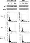
General cell cycle inhibition does not increase p73 levels. MDA-MB-231 and Rh30 cells were treated with 100 nM hydroxyurea (Hu) or 500 μM mimosine (Mim) or the appropriate vehicle control (C) for 24 h. Protein lysates were harvested and analyzed for p73 and actin by Western blotting, and cells were analyzed by flow cytometry to assess cell cycle profile.
FIG. 6.
FIG. 6.
Differential regulation of p53 family members by rapamycin. HMECs were treated with vehicle control (C) or rapamcyin for 12 h. Protein lysates were analyzed by Western blotting for p53, p63, p73, actin, pS6, and total S6. Results are representative of three independent experiments.
FIG. 7.
FIG. 7.
mTOR regulates p73 levels and activity. (A) mTOR knockdown induces p73. MDA-MB-231 cells were transduced with lentivirus engineered to express shRNA against the FRAP1 subunit of mTOR or with control lentivirus. Protein lysates were harvested 3 days after transduction, and mTOR, p73, p4EBP1, and actin were analyzed by Western blotting to demonstrate knockdown of mTOR levels and activity and induction of p73 levels. Western blots are representative of at least three independent experiments. (B) MDA-MB-231 cells were transduced with lentivirus engineered to express shRNA against GFP or p73. Protein lysates were harvested 5 days after transduction, and reduction of p73 levels was confirmed by Western blotting. (C) p73 activity is induced in MDA-MB-231 cells treated with 20 nM rapamycin, with or without concurrent serum starvation, and the result was verified using p73 RNAi. Cells were serum starved by preincubation in serum-free medium overnight before treatment with rapamycin. p73 RNAi was performed by transducing cells with lentivirus engineered to express shRNA against either GFP or p73 72 h before treatment. Total RNA was purified 48 h after treatment and reverse transcribed, and quantitative real-time PCR was performed with primers for the indicated genes. The samples were normalized to GAPDH, and the results are presented as increase over vehicle control values for an average of three experiments. Samples that exhibited a 30% or greater increase relative to control are indicated in red. Twelve of 17 genes exhibited a p73-dependent increase in RNA levels after rapamycin treatment and serum starvation. (D) Change in RNA as in panel C for INSR, TSC1, and XDH shows rapamycin/serum starvation-induced changes that are p73 dependent. Error bars represent standard deviations for three experiments. (E) Semiquantitative ChIP was performed to assess levels of p73 binding to genomic regions in INSR, TSC1, and XDH promoters or introns in Rh30 cells treated with vehicle or 40 nM rapamycin for 24 h. (F) MDA-MB-231 cells were transduced with shRNA lentivirus as for panel B and treated with 20 nM rapamycin or vehicle. At the indicated times cells from treated or control cultures were counted, and changes in cell growth rates due to p73 and rapamycin are shown. Error bars represent standard deviations from three experiments.
FIG. 8.
FIG. 8.
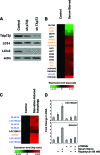
Analysis of p73-regulated genes in profiling studies of starvation and starvation-induced autophagy. (A) p73β knockdown decreases levels of autophagy markers. MDA-MB-231 cells were transduced with lentivirus engineered to express shRNA against TAp73 isoforms or against p73β isoforms. Protein lysates were prepared, and p73, actin, and the autophagy markers LC3-I and LC3-II were detected by Western blotting. (B and C) Genes from the p73 signature were assessed using publicly available data sets in which T98G glioblastoma cells were grown asynchronously or serum starved for 3 days before RNA harvest and microarray analysis (15) (B) or in which Awells B-lymphoblastoid cells were serum starved for 6 h or 24 h, inducing autophagy (25) (C). Known p53, p63, and/or p73 target genes indicated in orange are DDIT4 (31), DDB2 (53), DFNA5 (66), CDKN1C (10), GADD45A (49), JAG1 (88), and SESN2 (13). Cell lines are arranged in columns, grouped by treatment as indicated. Genes (annotated from Affymetrix probes) are in rows that are ordered based on hierarchical clustering results (data not shown). Color range shown indicates baseline transformed expression level on a log scale. Genes that are upregulated during autophagy or serum starvation that were selected for additional analysis are in blue. (D) Changes in RNA as in Fig. 4 for KLHL24 and LOC153222 indicate increases in RNA levels after rapamycin treatment in a p73-dependent manner in MDA-MB-231 cells.

"VSports最新版本" References

    1. Akerblad, P., R. Mansson, A. Lagergren, S. Westerlund, B. Basta, U. Lind, A. Thelin, R. Gisler, D. Liberg, S. Nelander, K. Bamberg, and M. Sigvardsson. 2005. Gene expression analysis suggests that EBF-1 and PPARgamma2 induce adipogenesis of NIH-3T3 cells with similar efficiency and kinetics. Physiol. Genomics 23206-216. - PubMed
    1. Amin, A. R., R. K. Paul, V. S. Thakur, and M. L. Agarwal. 2007. A novel role for p73 in the regulation of Akt-Foxo1a-Bim signaling and apoptosis induced by the plant lectin, concanavalin A. Cancer Res. 675617-5621. - PubMed
    1. Anderson, R. F., K. B. Patel, K. Reghebi, and S. A. Hill. 1989. Conversion of xanthine dehydrogenase to xanthine oxidase as a possible marker for hypoxia in tumours and normal tissues. Br. J. Cancer 60193-197. - PMC - PubMed
    1. Apweiler, R., T. K. Attwood, A. Bairoch, A. Bateman, E. Birney, M. Biswas, P. Bucher, L. Cerutti, F. Corpet, M. D. Croning, R. Durbin, L. Falquet, W. Fleischmann, J. Gouzy, H. Hermjakob, N. Hulo, I. Jonassen, D. Kahn, A. Kanapin, Y. Karavidopoulou, R. Lopez, B. Marx, N. J. Mulder, T. M. Oinn, M. Pagni, F. Servant, C. J. Sigrist, and E. M. Zdobnov. 2000. InterPro—an integrated documentation resource for protein families, domains and functional sites. Bioinformatics 161145-1150. - PubMed
    1. Avruch, J., Y. Lin, X. Long, S. Murthy, and S. Ortiz-Vega. 2005. Recent advances in the regulation of the TOR pathway by insulin and nutrients. Curr. Opin. Clin. Nutr. Metab. Care 867-72. - PubMed

"V体育官网入口" Publication types

"VSports手机版" MeSH terms