Skip to main page content
U.S. flag

An official website of the United States government

Dot gov

The . gov means it’s official. Federal government websites often end in . gov or . mil VSports app下载. Before sharing sensitive information, make sure you’re on a federal government site. .

Https

The site is secure. The https:// ensures that you are connecting to the official website and that any information you provide is encrypted and transmitted securely V体育官网. .

. 2008 Jun 5;2(6):576-83.
doi: 10.1016/j.stem.2008.03.009.

PUMA regulates intestinal progenitor cell radiosensitivity and gastrointestinal syndrome

Affiliations

PUMA regulates intestinal progenitor cell radiosensitivity and gastrointestinal syndrome

Wei Qiu et al. Cell Stem Cell. .

Abstract

Radiation is one of the most effective cancer treatments. However, gastrointestinal (GI) syndrome is a major limiting factor in abdominal and pelvic radiotherapy. The loss of crypt stem cells or villus endothelial cells has been suggested to be responsible for radiation-induced intestinal damage. We report here a critical role of the BH3-only protein p53 upregulated modulator of apoptosis (PUMA) in the radiosensitivity of intestinal epithelium and pathogenesis of GI syndrome. PUMA was induced in a p53-dependent manner and mediated radiation-induced apoptosis via the mitochondrial pathway in the intestinal mucosa VSports手机版. PUMA-deficient mice exhibited blocked apoptosis in the intestinal progenitor and stem cells, enhanced crypt proliferation and regeneration, and prolonged survival following lethal doses of radiation. Unexpectedly, PUMA deficiency had little effect on radiation-induced intestinal endothelial apoptosis. Suppressing PUMA expression by antisense oligonucleotides provided significant intestinal radioprotection. Therefore, PUMA-mediated apoptosis in the progenitor and stem cell compartments is crucial for radiation-induced intestinal damage. .


Figures
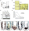
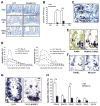
Figure 1
Figure 1. Radiation Induced Intestinal PUMA Expression
(A) PUMA mRNA expression in the jejunal mucosa of mice following whole body radiation (WRB) was evaluated by quantitative real-time RT-PCR. Values are means ± SD; n = 3 in each group. *p < 0.01; **p < 0.001. (B) PUMA, p53, and p21 protein expression in the jejunal mucosa of mice with indicated genotypes was determined by western blotting. β-actin was used as the control for loading. (C) PUMA mRNA in situ hybridization (ISH) in the jejunum of mice following radiation (magnification ×400). The selected area is shown at a higher magnification. Arrows indicate examples of PUMA-expressing cells. (D) PUMA mRNA expression (ISH) in the crypts was scored according to the cell position, pooled from 2 mice in each group. (E) PUMA expression in the columnar cells at the crypt base (CBCs) at 4 hr after 18 Gy. Examples of the CBCs are circled in red and stained positive for Ki-67 but negative for Paneth cell markers cryptdin 4 and MMP-7, magnification ×600.
Figure 2
Figure 2. PUMA Mediated Radiation-Induced Apoptosis in the Intestinal Progenitor and Stem Cells
(A) Apoptosis in the small intestinal crypts at 4 hr and 24 hr after 18 Gy WBR was assessed by TUNEL staining (brown), magnification ×400. (B) Apoptotic index in the crypts measured by TUNEL staining. Values are means ± SD; n = 3 in each group. *p < 0.005. (C) Example of apoptotic cells and their position in the crypt at 4 hr following 18 Gy WBR, magnification ×1000. Paneth cells are indicated with arrowheads while non-Paneth cells are indicated by arrows. (D) Apoptotic index at 4 hr (left panel) and 24 hr (right panel) following radiation. The apoptotic index was scored as the mean percentage of apoptotic cells of each cell position, pooled from four mice in each group. (E) PUMA ISH, and PUMA ISH/TUNEL double staining in the crypts 4 hr after 15 Gy, magnification ×400. Arrows indicate double-positive cells. (F) Serial sections (WT mice, 15 Gy at 4 hr) were subjected to TUNEL staining and Musashi IHC, magnification ×1000. Arrows indicate positive signals. (G) Radiation-induced apoptosis in the CBCs. Sections were stained with TUNEL or TUNEL followed by MMP-7 IHC with several CBCs circled, magnification ×600. (H) The fractions of crypts with at least one TUNEL-positive CBCs were calculated by counting 100 crypts with well preserved Paneth cell areas. Values are means ± SD; n = 3 in each group. *p < 0.005.
Figure 3
Figure 3. PUMA Did Not Contribute to Radiation-Induced Intestinal Endothelial Apoptosis
(A) Apoptosis induced by radiation in the villus was assessed by TUNEL staining (brown), magnification ×400. (B) Apoptotic index in the villus submucosa. Values are means ± SD. n = 3 mice in each group. (C) PUMA mRNA (red) ISH in the jejunal villi, magnification ×200. (D and E) The sections were double stained with TUNEL (brown) and CD105 (blue), magnifications ×200 and ×1000, respectively. Arrows indicate double positive cells. (F) The average fraction (%) of TUNEL+/CD105+ cells in the villus. Values are means ± SD; n = 3 mice in each group. (G) The average number of CD105+ cells in the villus. Values are means ± SD; n = 3 mice in each group. (H) Apoptotic index of CD105+ cells was determined at indicated times in WT and PUMA KO mice as in (F). (I) The number of CD105+ cells were determined at indicated times as in (G). (J) Apoptotic index of CD105+/PUMA+ cells in WT mice and that of CD105+ cells in PUMA KO mice were determined in 100 villi/mouse. Values are means ± SD; n = 3 mice in each group.
Figure 4
Figure 4. PUMA Mediated Radiation-Induced Intestinal Apoptosis via the Mitochondrial Pathway
(A) Bax, Bak, and cytochrome c (Cyto c) were analyzed in the cytosolic and mitochondrial fractions of intestinal mucosa by western blotting. β-actin and CoxIV were used as the controls for loading and fractionation. (B) Formation of Bax and Bak multimers was analyzed in the chemical cross-linker DSP-treated mitochondrial fractions by western blotting under nondenaturing conditions. (C) Caspase 3 activity was measured in the intestinal mucosa extracts. Values are means ± SD; n = 6 mice in each group. *p < 0.01. (D) Caspase 3 processing and β-actin levels were analyzed by western blotting. Results of two representative animals from the treated groups are shown. (E) Apoptotic index in the crypt was quantitated in WT and BAX KO mice after 15 Gy as in Figure 2B. Values are means ± SD; n = 3 mice in each group. W, WT mice; K, PUMA KO mice.
Figure 5
Figure 5. PUMA Deficiency Prolonged the Survival of Mice Following WBR
(A) BrdU incorporation index in the small intestine was quantitated by counting 100 crypts. Values are means ± SD; n = 3 mice in each group. (B) Crypt regeneration was calculated by counting 10 cross-sections following BrdU staining. Values are means ± SD; n = 4 in each group. (C) Regenerated crypts were quantitated in WT and BAX KO mice after 15 Gy WBR as in (B); n = 3 in each group. (D) Survival curves of mice subjected to 15 Gy or 18 Gy WBR. (E) H&E staining of the small intestine sections, magnification ×200.
Figure 6
Figure 6. PUMA Mediated p53-Dependent Crypt Cell Apoptosis Induced by Radiation
(A) PUMA mRNA expression in the intestinal mucosa was evaluated by quantitative real-time RT-PCR. Values are means ± SD; n = 3 in each group. *p < 0.001; **p < 0.01. (B) PUMA, p21, p53, and β-actin protein expression in the intestinal mucosa were determined by western blotting. (C) Apoptotic index measured by TUNEL staining. Values are means ± SD, and n = 3 in each group. *p < 0.01, **p < 0.001. (D) Regenerated crypts were quantitated after 15 Gy WBR by counting 10 BrdU stained sections. Values are means ± SD; n = 4 in each group.
Figure 7
Figure 7. PUMA Antisense Oligonucleotides Suppressed Crypt Apoptosis and Prolonged Survival of Mice Following WBR
(A) The effects of PUMA antisense oligonucleotides on PUMA expression in MEFs after Brefeldin A (20 nM) treatment were evaluated by western blotting. (B) PUMA expression in the small intestinal mucosa of mice treated with PUMA sense or antisense (AS) oligonucleotides at 24 hr following 15 Gy WBR. (C and D) Apoptotic and BrdU incorporation indices in the crypt 24 hr after 15 Gy WBR. Values are means ± SD; n = 3 mice in each group. (E) Crypt regeneration at 96 hr following 15 Gy WBR. Values are means ± SD; n = 5 mice in each group. (F) Survival curves of mice following 15 Gy WBR.

VSports app下载 - Comment in

References

    1. Barker N, van Es JH, Kuipers J, Kujala P, van den Born M, Cozijnsen M, Haegebarth A, Korving J, Begthel H, Peters PJ, Clevers H. Identification of stem cells in small intestine and colon by marker gene Lgr5. Nature. 2007;449:1003–1007. - PubMed
    1. Booth D, Potten CS. Protection against mucosal injury by growth factors and cytokines. J Natl Cancer Inst Monogr. 2001;29:16–20. - PubMed
    1. Carson-Walter EB, Hampton J, Shue E, Geynisman DM, Pillai PK, Sathanoori R, Madden SL, Hamilton RL, Walter KA. Plasma-lemmal vesicle associated protein-1 is a novel marker implicated in brain tumor angiogenesis. Clin Cancer Res. 2005;11:7643–7650. - VSports注册入口 - PubMed
    1. Ch’ang HJ, Maj JG, Paris F, Xing HR, Zhang J, Truman JP, Cardon-Cardo C, Haimovitz-Friedman A, Kolesnick R, Fuks Z. ATM regulates target switching to escalating doses of radiation in the intestines. Nat Med. 2005;11:484–490. - PubMed (V体育安卓版)
    1. Erlacher M, Michalak EM, Kelly PN, Labi V, Niederegger H, Coultas L, Adams JM, Strasser A, Villunger A. BH3-only proteins Puma and Bim are rate-limiting for gamma-radiation- and glucocorticoid-induced apoptosis of lymphoid cells in vivo. Blood. 2005;106:4131–4138. - PMC - PubMed

MeSH terms

Substances

"V体育ios版" LinkOut - more resources