VSports最新版本 - Skip to main page content
U.S. flag

An official website of the United States government

Dot gov

The . gov means it’s official. Federal government websites often end in . gov or VSports app下载. mil. Before sharing sensitive information, make sure you’re on a federal government site. .

Https

The site is secure V体育官网. The https:// ensures that you are connecting to the official website and that any information you provide is encrypted and transmitted securely. .

. 2008 May 15;68(10):3835-43.
doi: 10.1158/0008-5472.CAN-08-0215.

V体育官网 - An anti-transforming growth factor beta antibody suppresses metastasis via cooperative effects on multiple cell compartments

Affiliations

An anti-transforming growth factor beta antibody suppresses metastasis via cooperative effects on multiple cell compartments

Jeong-Seok Nam et al. Cancer Res. .

Abstract

Overexpression of transforming growth factor beta (TGF-beta) is frequently associated with metastasis and poor prognosis, and TGF-beta antagonism has been shown to prevent metastasis in preclinical models with surprisingly little toxicity. Here, we have used the transplantable 4T1 model of metastatic breast cancer to address underlying mechanisms VSports手机版. We showed that efficacy of the anti-TGF-beta antibody 1D11 in suppressing metastasis was dependent on a synergistic combination of effects on both the tumor parenchyma and microenvironment. The main outcome was a highly significant enhancement of the CD8+ T-cell-mediated antitumor immune response, but effects on the innate immune response and on angiogenesis also contributed to efficacy. Treatment with 1D11 increased infiltration of natural killer cells and T cells at the metastatic site, and enhanced expression of coactivators (NKG2D) and cytotoxic effectors (perforin and granzyme B) on CD8+ T cells. On the tumor cells, increased expression of an NKG2D ligand (Rae1gamma) and of a death receptor (TNFRSF1A) contributed to enhanced immune cell-mediated recognition and lysis. The data suggest that elevated TGF-beta expression in the tumor microenvironment modulates a complex web of intercellular interactions that aggregately promote metastasis and progression. TGF-beta antibodies reverse this effect, and the absence of a major effect of TGF-beta antagonism on any one cell compartment may be critical for a good therapeutic window and the avoidance of autoimmune complications. .


Figures
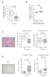
Figure 1
Figure 1. Suppression of lung metastasis by 1D11 in the 4T1 model is associated with increased infitration of T-cells and reduced angiogenesis
A. Numbers of histologically detectable metastases in the lung. 4T1 cells were implanted orthotopically into the left thoracic mammary fat pad of syngeneic BALB/c mice and mice were randomized to treatment with anti-TGF-β (1D11) or control 13C4 antibody (CON) antibody starting at day 1 after innoculation. Primary tumors were surgically excised at day 10, mice were euthanized on day 28. Boxes, median values with upper and lower quartiles; whiskers, range. (Control group, 11 mice; 1D11 group, 13 mice). B. Comparison of 1D11 efficacy in reducing lung metastasis number using either the orthotopic implantation or the tail vein injection experimental formats. The extent of metastasis suppression for each experiment was determined by comparing the median number of metastases in the 1D11-treated group with that in the control group. Each point represents the results of an independent experiment that contained a minimum of 10 mice/treatment group. Metastasis suppression was statistically significant in each case. C and D. The effect of 1D11 on microvessel density (C: CD31 marker) or on the number of infiltrating T-cells (D: CD3 marker) in the primary tumors and lung metastases from the experiment in (A) was determined by semiautomated analysis of immunohistochemically stained sections as described in Methods. Representative images are shown for each marker. A minimum of 10 randomly selected fields were quantitated for each primary tumor, or the entire area of every metastasis in each set of lung lobe cross-sections was evaluated, to determine the mean marker density for that mouse. For primary tumors, 8 mice were evaluated/group and for metastases, 8-11 mice were evaluated/group. Boxes, median values with upper and lower quartiles; whiskers, range.
Figure 2
Figure 2. Maximum efficacy of 1D11 in suppressing metastasis is dependent on the presence of CD8+ cells, NK cells and an intact TGF-β response in the tumor cell
4T1 cells were injected into the tail-vein of BALB/c mice, and mice were treated with 1D11 or CON antibody with dosing and scheduling as in Fig. 1. Mice were euthanized on day 21 and the number of grossly visible metastases/lung was determined. All experimental groups had 10-13 mice/group. Boxes, median values with upper and lower quartiles; whiskers, range. A. Effect of CD8 cell depletion. Where indicated, mice were treated with anti-CD8 antibody, prior to the injection of tumor cells, to deplete CD8+ cells. One experiment representative of two replicates is shown. B. Effect of NK cell depletion. Where indicated, mice were treated with anti-asialoGM1, prior to injection of tumor cells, to deplete NK cells. C. Effect of loss of TGF-β response in the tumor cell. 4T1 cells were transfected with either an empty vector (EV) or with a dominant negative type II TGF-β receptor (DNR) and pooled transfectants were used for the metastasis assay. D. Summary of the efficacy of 1D11 in suppressing metastasis following the interventions above. The extent of suppression of lung metastases by 1D11 was first determined by comparing median numbers of lung metastases in mice treated with 1D11 and CON for each of the individual interventions. The efficacy of 1D11 following each experimental intervention was then expressed as a % of the maximum therapeutic effect seen in the unmanipulated (“intact”) system. Results are the mean +/- SD for the indicated number of replicate experiments.
Figure 3
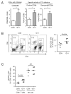
Figure 3. Effect of 1D11 treatment on numbers of immune cells and markers of activity
A. RTQ-PCR analysis of mRNA levels of CD8b, and of the cytotoxic T-cell effectors, perforin and granzyme B from primary tumors treated with anti-TGF-β (1D11) or control (CON) antibody. Results are the mean +/- SD for 5 tumors/treatment group. P<0.05. B and C. FACS analysis of immune cells recovered from tumor-bearing lungs of mice injected orthotopically with 4T1 cells and treated with anti-TGF-β (1D11) or control (CON) antibody. Lungs were harvested at day 28, digested with collagenase, enriched for viable cells using a Percoll gradient, and then immune cells were analyzed for expression of specific markers by FACS. B. Representative FACS plot for determination of the effect of antibody treatment on the representation of T-cells (CD3+) or NK cells (CD3-DX5+) in the leukocyte gate by dual immunostaining. The scatter plot shows the % NK cells in the leukocyte gate for 5 mice/treatment group. Median values are indicated. C. The % of cells expressing the activator/coactivator NKG2D was determined individually for the CD8+ and the DX5+ (NK) cell populations for 5 mice/group. Median values are indicated.
Figure 4
Figure 4. Efficacy of 1D11 involves effects on the tumor cell as well as the immune compartment
A. Effect of TGF-β blockade on metastatic efficiency in the presence or absence of CD8+ cells. TGF-β response was blockaded specifically in the tumor cell by transfection with a dominant negative type II receptor (DNR) or empty vector control (EV). The number of lung metastases was determined using the tail vein injection protocol, with or without pre-treatment of the mice with anti-CD8 antibody to deplete CD8+ cells (12 mice/group). The horizontal line indicates the median number of metastases. B. RTQ-PCR and FACS analysis of Rae-1γ expression in 4T1 cells after 4 days of treatment with TGF-β (5ng/ml). Rae-1γ mRNA was normalized to 28SrRNA, and results are the mean +/- SD (n=3). C. RTQ-PCR and FACS analysis of Rae-1γ expression in primary tumors derived from mice treated with anti-TGF-β (1D11) or control antibody (CON). RTQ-PCR results are the mean +/- SD for 5 tumors/group and Rae-1γ mRNA expression was normalized to 28s rRNA. FACS results for two representative tumors in each treatment group are shown. *P<0.001; Students T-test.
Figure 5
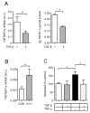
Figure 5. Effect of TGF-β on TNF-α receptor expression and sensitivity of 4T1 cells to induction of apoptosis by TNF-α
A. Expression of TNF-α receptor 1 (TNFRSF1a) mRNA and protein in 4T1 cells were treated for 2 days in vitro with 5ng/ml TGF-β or vehicle control. TNFRSF1a mRNA was determined by RTQ-PCR, and the level of TNFR1 protein in 4T1 cell lysates was detected by ELISA assay. Results are the mean +/- SD for 3 determinations. B. RTQ-PCR analysis of TNFRSF1a expression in 4T1 primary tumors following 9 days of treatment in vivo with anti-TGF-β (1D11) or control (CON) antibody. TNFRSF1a expression was normalized to the 28S rRNA in each case. Results are mean +/- SD for 5 tumors/treatment group. C. In vitro apoptosis assay in 4T1 cells after treatment with TGF-β (5ng/ml) or vehicle control for 4 days, with TNF-α (25ng/ml) also present for the final two days. Results are normalized to the no TGF-β, no TNF-α control conditions, and are the mean +/- SD of 3 determinations. * indicates P<0.001, Student's T-test.
Figure 6
Figure 6. Model for the multiple effects of anti-TGF-β antibody on different cellular compartments within the tumor
Antagonism of TGF-β by anti-TGF-β antibody treatment in the 4T1 breast cancer model has many small magnitude effects locally on the tumor parenchyma and stromal compartments. Most of these effects cooperate to enhance effective anti-tumor immune responses, and aggregately they result in a reduction in metastatic burden (“death by a thousand cuts”). Some effects lead to synergy between different cellular compartments, such as the upregulation of Rae-γ, an NKG2D ligand, on the tumor cell and enhanced expression of NKG2D on CD8+ T-cells. *The effects of 1D11 on Bsp1, IL-6 and IL-17 expression in this model are been described elsewhere ((17) and accompanying manuscript)

References

    1. Pardali K, Moustakas A. Actions of TGF-β as tumor suppressor and pro-metastatic factor in human cancer. Biochim Biophys Acta. 2007;1775:21–62. - PubMed
    1. Derynck R, Akhurst RJ, Balmain A. TGF-β signaling in tumor suppression and cancer progression. Nat Genet. 2001;29:117–29. - PubMed
    1. Gold LI. The role for transforming growth factor-β (TGF-β) in human cancer. Crit Rev Oncog. 1999;10:303–60. - PubMed
    1. Teicher BA. Transforming Growth Factor-β and the Immune Response to Malignant Disease. Clin Cancer Res. 2007;13:6247–51. - "VSports最新版本" PubMed
    1. Bierie B, Moses HL. Tumour microenvironment: TGFβ: the molecular Jekyll and Hyde of cancer. Nat Rev Cancer. 2006;6:506–20. - PubMed

Publication types